Browsed by
Category: Featured

Creativity and Stress

Creativity and Stress

Written by Quinn Klessel  –  Artwork by Connie Ulm  –  July 26, 2021

According to the American Psychology Association (2019), roughly 75% of Americans report that they experience physical, mental, and behavioral symptoms from an excess of stress. Biologically, stress can be defined as, “a condition that imposes severe demands on the physical and psychological defenses of the organism”(APA Dictionary of Psychology, 2021). The human body has a fine tuned way of dealing with situations that the individual deems to be stressful. The issue is that when we encounter too many incidents that we deem stressors, we overuse this stress response and our bodies experience negative side effects. Too much stress has the potential to lead to a decrease in productivity, but there are ways to counteract this. One way is through participating in creative arts, which has the ability to lower stress levels and increase both productivity and problem solving abilities. A stressed out person may respond to the advice of “Take a break, draw a picture” by saying, “That is a waste of my time.” However, one would find that this person would have a much less grumpy response after they took a break and drew a picture. Their stress would be lowered. Don’t believe me? At the end of this article, if you are experiencing some stress (no matter how much or how little), try your hand at the creative activity I shall put forth.

When a person identifies something as a stressor, the brain registers a distress signal that causes a fight, flight, or freeze response (Katz et al., 2021). The body releases a hormone called epinephrine during this response, and this hormone triggers a series of biological changes in our bodies that allow us to more efficiently deal with danger. If our brain continues to perceive danger for a prolonged period of time, it uses a chain reaction within a system called the hypothalamic-pituitary-adrenal axis (HPA axis) to release cortisol, a hormone into our system. This system to deal with stress works perfectly under most circumstances. However, if our brain reacts to situations in a way in which we perceive extreme danger where there is not, we can overuse this stress response and cause harm to our bodies from an excess of the secretion of glucocorticoids, such as cortisol (Chu et al., 2021). The negative side effects of excess stress include headache, chest pain, nausea, anxiety, restlessness, irritability, and depression (MayoClinic, 2021). These are all things that at the least have the potential to inconvenience an individual’s life.

One might wonder at this point, is stress just entirely harmful? Should humans try to experience as little stress as possible as to avoid all risk of the negative biological effects of too much stress? While too much stress can be harmful to our bodies, too little stress is also not ideal. It was once thought that glucocorticoids, like cortisol, only inhibited brain functions. This would imply that stress only inhibited brain function. However, the Yerkes-Dodson law presents a different theory. This law showcases a graph containing an inverted-U shape with the x-axis being arousal and the y-axis being performance. According to this law, there is an optimal amount of stress needed for peak performance; too little or too much stress will not cause this best scenario. 

Many studies have been conducted using the injection of glucocorticoids into mice and other organisms to see if the theory that stress hormones only inhibit cognitive function is true. While conducting these tests, it was discovered that there are two types of glucocorticoid receptors in the brain: Type 1 and Type 2. At a state of rest, glucocorticoids are mostly bonded to the Type 1 receptor. However, when more glucocorticoids are injected to the point where the Type 1 to Type 2 receptor bonding ratio is approximately equal, it is found that memory function is at its peak. Therefore, when the glucocorticoids have bonded to Type 2 receptors as much as they have bonded to Type 1 receptors, the mice show the best memory performance. This demonstrates the idea that an inverted-U graph with the x-axis representing the amount of glucocorticoids injected and the y-axis representing memory function is the ideal representation. Too little or too much of the glucocorticoids will not provide peak memory function, thus, too little or too much stress will not provide peak memory function (Lupien et al., 2007).


The conclusions that can be drawn from this experiment are that some stress is helpful to increase cognitive function, however too much stress may be detrimental. Now that we understand stress is not an entirely bad thing, we understand that we should not be making an effort to remove humans from stressful situations all together, but should be making an effort to reduce stress when there is an unhealthy amount. The strategies we use to reduce stress can be personalized based on how an individual experiences stress. The many facets of the creative arts are ideal for treatment personalization, and this is important because not all activities illicit the same biological response for all individuals.

Serotonin and dopamine are arguably the best hormones for encouraging a creative space (Shiv, 2014). Serotonin governs whether an individual feels calm and secure, or experiences anxiety and fear, and dopamine governs whether an individual feels excited and motivated, or bored and apathetic. The proper amount of serotonin and dopamine will allow a person to live in a calm and motivational environment, thus an environment that best promotes creativity (Underwood, 2014).  An increase in cortisol is known to reduce serotonin levels, potentially by increasing the expression of a serotonin transporter that elevates the uptake of serotonin (Tafet et al., 2001). Therefore, chronic stress has the ability to decrease one’s ability to live in a space of creativity.


Since the creative arts allow for treatment personalization, it is possible to find a creative arts activity that could increase serotonin and dopamine levels in many individuals. Some activities you could try out include doing yoga, drawing, creative writing, sculpting, writing music, playing music, dancing, etc. As every person is different and has individualized interests, not all of these activities will have the same effect on every person. Luckily, the creative arts allow for many options to choose from.


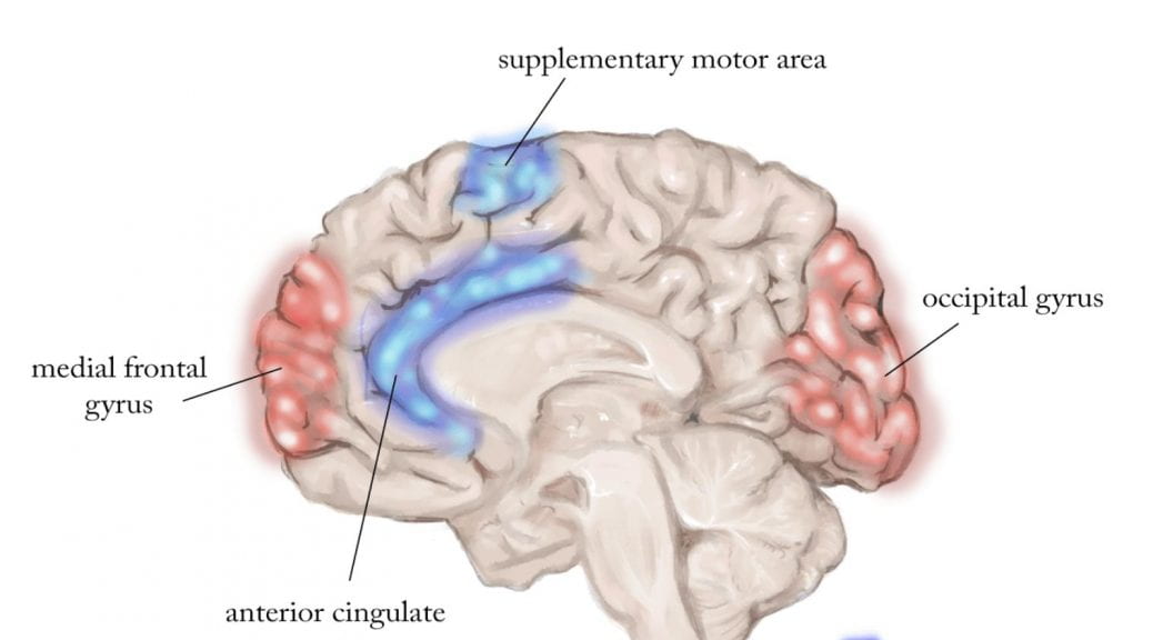
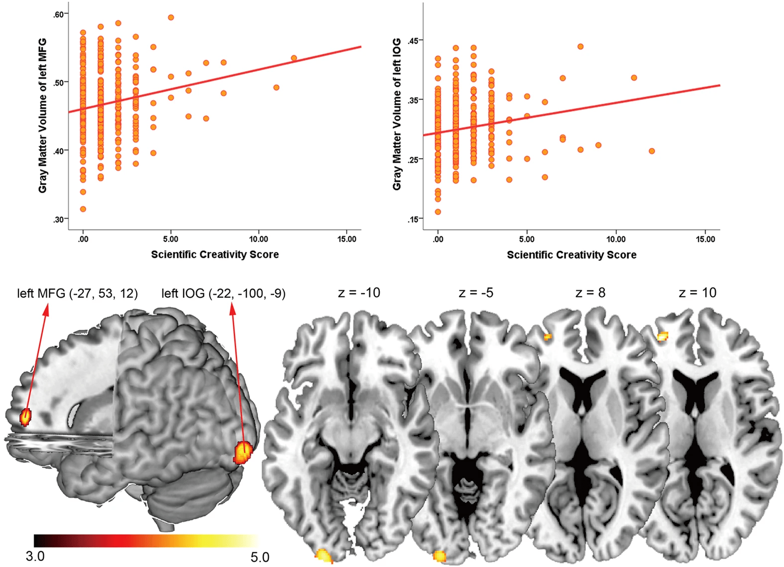
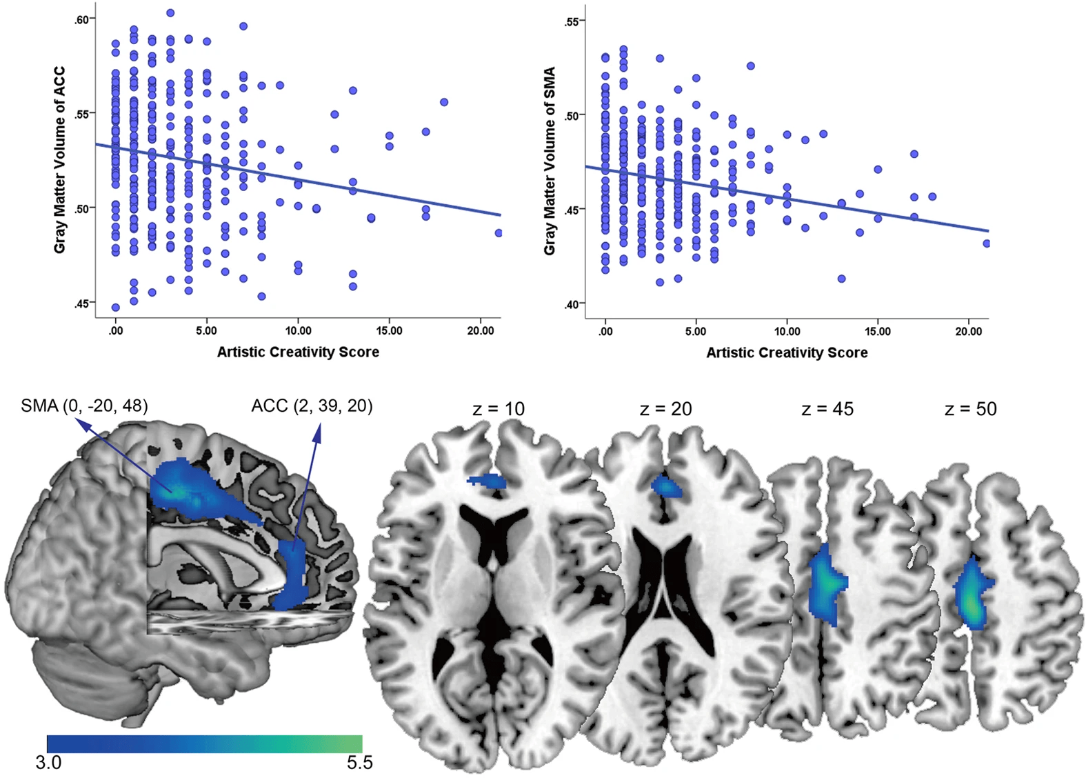
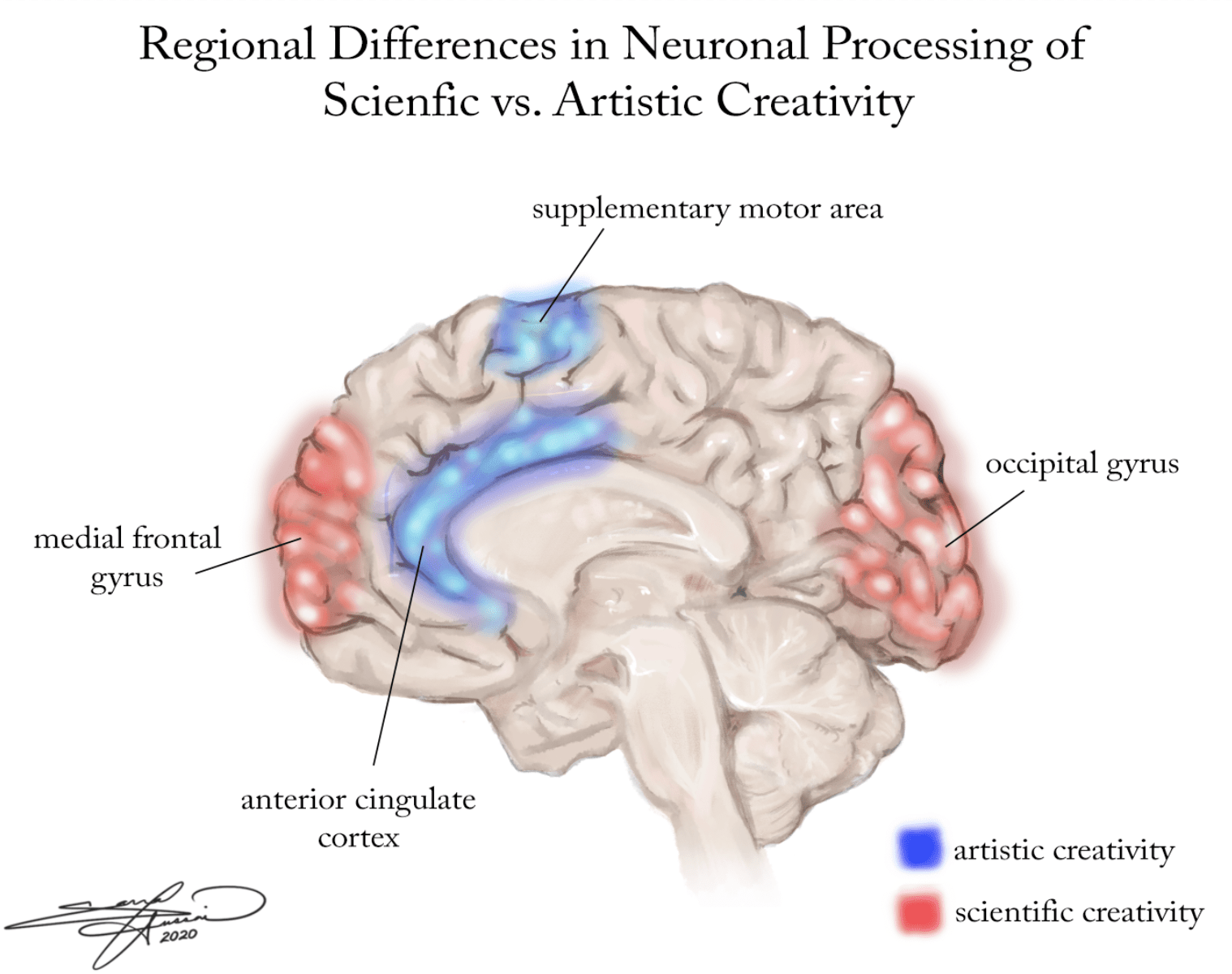
It is important to note that the process of creativity is executed in the same part of the brain as the process of problem solving: the frontal lobe (Kleibeuker et al., 2013). Increasing creativity can increase one’s ability to problem solve and thus increase one’s performance at a task. A human being cannot have an excess of stress hormones, such as cortisol, and be able to efficiently create hormones that increase creativity and problem solving, such as serotonin.  Thus, in addition to what is learned from glucocorticoids and memory in the Yerkes-Dodson law graph, we also know from an endocrinological standpoint why decreasing stress is important for peak performance. Performing creative arts activities will activate the frontal lobe, and thus activate the part of the brain that helps one problem solve and perform.


Studies have been done which show a significant decrease in cortisol levels after multiple sessions of participating in creating art. One study published in the Journal of the American Art Therapy Association (2016) showed 75% of participants had a significant decrease in cortisol levels after 45 minutes of creating art. Another study in the Journal of Nursing Education (2005) found that incorporating a creative arts activity into the classroom environment significantly reduced stress and anxiety levels amongst the nursing students. An experiment was also conducted with girls who were living in family-like centers (Kheibari et al., 2014). For one week, half of the girls participated in art therapy, while the other half did not. The study found a significant reduction in the level of anxiety amongst the girls who participated in the art therapy. The conclusion can be made from these studies and many more like them that engaging in the creative arts has the potential to be a tool for lowering stress levels and increasing the health of those managing chronic stress. We also know from the Yerkes-Dodson law that these conclusions suggest that engagement in the arts or in art therapy could increase the work performance of those who experience stress levels that are too high.


The introduction of creative arts intervention into both professional and academic settings could be beneficial to increasing the performance of both employees and students. From a neurological standpoint, creating art has proven to be significant in decreasing stress levels amongst individuals and thus has the ability to increase the overall performance of these individuals in a multitude of life tasks. The understanding of this concept and implementation of it has the potential to be a non-invasive and enjoyable solution to stress inhibiting work. Allowing for human beings to use creativity and operate in a creative space can improve lives drastically and this concept deserves more attention. If you would like to put the concepts I have put forward to the test, I suggest you try this quick and easy creativity inducing activity. Perhaps if you are currently feeling stressed, this activity will ease your mind.


Using only circles, squares, and triangles, draw a picture in response to this prompt: What was your biggest strength today?

This activity may lower your stress, or you may want to participate in an art form more specific to your liking. Regardless, keep your mind open, and allow it to fill all of the creative spaces it would like to.

Synesthesia – The Joining of the Senses

Synesthesia – The Joining of the Senses

Written by Anna Peris – Artwork by Anna Peris – July 9, 2021

Vincent Van Gogh, writer Vladimir Nabokov, and Australian singer Lorde, a seemingly unrelated trio, share one unifying experience – all are among the small group of people with the neurological condition of synesthesia. People with this condition, called synesthetes, involuntarily experience sensory reactions, from a stimulus that activates a different sensory pathway. Since the sensory experiences and the stimulus that causes them varies widely among synesthetes, over eighty forms of synesthesia have been identified. Two of the most commonly known types are grapheme-color synesthesia and chromesthesia. Grapheme-color synesthesia causes a person to perceive particular colors when seeing letters, and chromesthesia is when sounds, whether they are musical or ambient, prompt individuals to see colors. Some synesthetes experience words as having unique flavors, while, for others, sounds will elicit physical sensations. Many possess more than one type of synesthesia (Safran et al, 2015).  

The exact physiological processes behind synesthesia have not been fully revealed, but neurological research has demonstrated that sensory experiences are related to an abundance of connections in the white matter tracts between sensory regions of the brain. When a stimulus activates one sensory region, this heightened connectivity causes the neural activity to overflow into other regions. For grapheme-color synesthesia, the adjacent regions responsible are the fusiform gyrus and V4 regions. The fusiform gyrus is a region that deals with visual appearance and recognition, and the V4 region is a part of the visual cortex that deals with color perception. FMRI studies have indicated that the white matter connectivity for grapheme-color synesthetes is heightened in the superior parietal lobule, as shown in the image below (Hubbard et al, 2005).

For those synesthetes that associate numerical sequences and other concepts like months of the year with color, it is thought that this is a result of the interaction between the angular gyrus, a part of the inferior parietal lobule associated with numeric processing, and the V4 region. This conclusion of interaction between sensory regions has been solidly supported as the basis for these two forms of synesthesia, while there is less evidence that this process is what controls some of the more rare forms of the condition. Genetic studies of synesthetes have suggested that synesthesia can be genetically inherited. However, since the studies identified numerous genes inconsistently related to inheritance of synesthesia among different families, no particular gene or gene cluster has been labelled as the singular gene responsible for synesthesia inheritance (Ramachandran and Hubbard, 2001). 

While synesthesia is by no means common, it is less rare than commonly thought, with an estimated 2 to 4 percent of the global population experiencing some form of the condition. Among artists and musicians, it is more prevalent. Many of the famous examples of synesthetes fall into this group. Since many synesthetes constantly perceive such strong visual experiences due to sounds or letters, these colours and shapes often inspire works of art both visual and musical. Besides Van Gogh, some famous artists with this condition include Wassily Kandinsky (a Russian artist), Edgar Degas (a French artist), and Edvard Munch (a Norwegian artist). The influence of chromesthesia is highly evident in the works of Kandinsky. He often wrote about how he saw colors when he heard music, and he heard music when creating his artwork. In a 1915 letter to colleague Gabriele Munter, he wrote, 

First I will make different color tests: I will study the dark – deep blue, deep violet, deep dirty green, etc. Often I see the colors before my eyes. Sometimes I imitate with my lips the deep sounds of the trumpet – then I see various deep mixtures which the word is incapable of conceiving and which the palette can only feebly reproduce” (Boehmer, 2011). 

Kandinsky’s “Impression III,” inspired by a concert he attended

Melissa McCracken is an American contemporary artist with chromesthesia, and she creates paintings based on the colors particular songs evoke for her, as shown in a few of her works below. 

Melissa McCracken, Inspired by “Karma Police” by Radiohead

Melissa McCracken, Inspired by Bach’s Cello Suite No. 1

Beyond the world of visual art, many musical artists are synesthetes. Composers such as Franz Liszt, Jean Sibelius, and Nikolai Rimsy-Korsakov all experienced chromesthesia. Contemporary musicians with the condition include Jimi Hendrix, Lady Gaga, Frank Ocean, Pharrell Williams, and countless others. Some synesthetic songwriters have described what they see when hearing their own compositions. In an interview, Billy Joel mentioned that some of his more sentimental songs like “Vienna” are equated with soft shades of green and blue. For some musicians, perfect pitch overlaps with synesthesia, and scientific studies have demonstrated that these two phenomena often coincide. A 2013 study reported that out of 768 subjects with validated absolute pitch, 155 exhibited synesthesia, demonstrating a strong association between these two traits. Additionally, the study concluded that there was a significant genetic overlap between the two phenomena (Gregersen, et al., 2013). 


While the first written example of synesthesia is from Pythagoras in 500 BC, synesthesia was not formally discovered as a condition until the nineteenth century. Likely synesthetes prior to this time period, including Van Gogh, were often misunderstood as insane when relating their experiences to others. As more research was undertaken, and brain imaging validated the experiences reported by synesthetes, the condition was gradually destigmatized (Safran, 2015). Research into synesthesia continues today, as more and more hypotheses about the physiological and genetic bases of the condition are proposed. One such hypothesis that holds some popularity is that many individuals experience synesthesia to some degree, even if it is very slight, based on the observation that some people experience involuntary associations between senses in response to a few limited stimuli. Continued synesthesia research is thought to be helpful to our scientific understanding of other conditions such as autism, dyslexia, and loss of a particular sensory function. It may also contribute to research about memory and absolute pitch (Ramachandran and Brang, 2008). No matter how many discoveries about the condition are made, synesthesia still remains as a source of intrigue and an inspiration of artistic work. 

Creative Madness

Creative Madness

Written by Sheridan Scott – Artwork by Zena Meighan – June 19, 2021

 Have you ever been tempted to cave to temptation and slice off your ear? Or maybe  you’re dying to experience the robust flavor of yellow paint? Personally, I’ve only ever eaten yellow playdough, unlike our friend Vincent Van Gogh (a Dutch artist). Van Gogh was an irritable alcoholic, diagnosed with “a sort of epilepsy with hallucinations and episodes of agitation and confusion provoked by alcoholic excess” (Hemphill, 1961, p. 28). Arguably, his alcohol dependence stemmed from an inability to manage emotional extremities, as he once said, “If the storm within gets too loud, I take a glass more to stun myself” (Blumer, 2002). Despite his inner turbulence, Van Gogh created masterpieces that will continue to awe generations. Notably, he created more self-portraits than the average artist, several of which illustrate his distress and unease. His self-portrait that stands apart from the rest, however, showcases Van Gogh with a pipe and a bandaged ear. This portrait is unique not only for obvious ear-related reasons but also because, for once, he appears tranquil (Hemphill, 1961, p. 32).

Van Gogh is far from the only renowned artist who is also known for their madness. While there are several like him, Louis Wain (an English artist) sticks out most in my mind. Admittedly, it’s probably because of my similar obsession with cats. When Wain’s wife was diagnosed with breast cancer at a young age, he, naturally, bought her a lovely cat named Peter. Drawing cats, both to raise his wife’s spirits and manage his own feelings of despair, became his new obsession. His earlier drawings were more realistic representations, quite the opposite of his later. Wain’s life took a turn for the worse when his adored wife Emily died, later followed by his sister and adorable cat, Peter. As Wain’s life changed, so did his paintings. No longer realistic, his most recent paintings depict cats (still an obsession but can you blame him?) in elaborate, abstract forms. In later years, Wain suffered bouts of paranoia and psychosis and was institutionalized up until his death, although he never ceased painting his beloved cats (Damiani, S., & Fusar-Poli, L., 2018). 

Since so many great artists and writers suffer from mental illnesses, we are led to wonder whether the term “mad artist” is scientifically backed. Signs of tribulations and illness exude from masterpieces, but does this imply cognitive disinhibition improves artistic cognition? I present to you: the inverted U. Picture an arch that resembles the great American fast food chain,  McDonald’s original logo. The x-axis signifies the degree of top-down control, ranging from normal to defocused to impaired; and the y-axis represents someone’s degree of originality. Before jumping ahead, let’s talk about top-down control, particularly top-down dysfunction. Top-down control refers to how knowledge and expectations impact informational processing. Individuals who exhibit top-down dysfunction (fronto-striatal dysfunction) exhibit a variety of behaviors: poor inhibition, latent disinhibition, distractibility, and impulsivity. Connecting the dots, you notice these behaviors persist in a wide array of mental disorders, such as Attention Deficit Hyperactivity Disorder (ADHD), bipolar disorder, schizophrenia, substance abuse, and more (Abraham, 2014). Now, let’s re-examine that McDonald’s logo. Individuals who exhibit top-down dysfunction fall under the defocused or impaired part of the curve. What this means is that moderate levels of top-down dysfunction are associated with a far greater degree of creativity, while impaired or debilitating levels are associated with a below average degree of creativity (Abraham, 2014).

A key take-away: if you or a loved one struggles with top-down dysfunction, buy an easel or grab a pen. Let the “madness” make some magic.


The Neurobiological Understanding of Dance Movement Therapy

The Neurobiological Understanding of Dance Movement Therapy

By Tracy Zhang  –  Artwork by Connie Ulm  –  June 19, 2021

Picture this scenario: you are in the car listening to the radio after a long day of work sitting in the office. Your favorite song comes on, and you start tapping your fingers to the beat, feeling the music flow through your veins. As the song escalates towards the climax, you find your arms, feet and legs grooving to the beat of the music as well. While the movements may seem like a natural response to the rhythm of music, several regions of the brain, such as the motor cortex, somatosensory cortex, basal ganglia, and cerebellum are at play.


Dance movement therapy is a form of art therapy that incorporates the use of movement and emotions. It is rooted in the understanding that the body and mind are interconnected: the movement of the body can be used to understand a person’s mental and emotional state. Further explained by the American Dance Therapy Association, “movement promotes emotional, social, cognitive and physical integration of the individual”. Dance movement therapy can be conducted in sessions with individuals, families, couples and in groups. There are many benefits to dance movement therapy because it can be applied to help with a variety of issues. These issues vary  from physical and mental health issues as well as social and cognitive issues. The benefits include increased coordination and mobility, building positive body image and self esteem and improved communication skills (Good Therapy).  In addition, people with complex traumas, “severe events that tend to be chronic and undermine a child’s personality development and fundamental trust in relationships” (Kliethermes et al, 2014), often develop defense mechanisms preventing them from expressing their emotions verbally. Dance movement therapy provides people with an outlet to understand their own emotions and experience positive growth. Dance movement therapy also plays a role in certain neurological functionings such as arousal/rest, memory and the mirror neuron system. The neurobiology of dance movement therapy can help one understand and highlight this connection of the body and mind.


Dance movement therapy sessions do not have a standardized structure and each therapist has their own style of running their sessions. However, there are main principles and techniques most dance therapists incorporate into their sessions that were developed by the pioneers of Dance Movement Therapy which were Marian Chace, Mary Whitehouse, and Trudy Schoop. Therapy sessions are typically conducted in phases: warmup, theme development and closure (Nemetz, 2005). The purpose of warmup is to get the clients familiar and comfortable with each other and the therapist. The warm up includes initial contact techniques (mirroring the instructor, clarifying expressive movement and implementing verbal and nonverbal contact to extract movement responses), group development formation and group expression. For instance, a client with emotional distress may contract their body and hunch forward in a way that shows they are afraid. A therapist may then continue that movement with a relaxed action of expanding the body (Nemetz, 2005). Soon, the group becomes more cohesive as a unit and develops group trust through simple rhythmic movements and voice narration. This leads the session onto the theme development phase. In this phase, the therapist would lead the group through an exploration of the themes identified in the warm up using imagery, verbalization and movement. For instance, as the group does a simple side to side arm swing together, one client may display a different variation of this movement subconsciously with a deeper meaning behind it. As a result, the therapist explores this by either asking questions or intensifying that movement or making noises that she feels while perceiving that variation.  Lastly, the session ends with closure where the clients form a circle engage in repetitive group movements, allowing them to leave with a feeling of emotional support and connection (Nemetz, 2005). These group movements may include swinging their arms while holding hands or raising their hands high together.

Dance movement therapy is an effective form of therapy and its methods are understood through a neurological lens. Initial approaches to dance movement therapy are effective in guiding clients to better understand their body. These methods such as rhythm breathing bring awareness to the senses experienced by the body and induce a state of relaxation. The feeling of safety and balance will allow them to process larger and more complex emotions. This can be understood through one of the cranial nerves in our brain, the vagus nerve (Homann, 2010). The vagus nerve is located at the medulla (the lower portion of the brainstem) and travels down the spine connecting the heart, lungs, stomach, and intestines. The vagus nerve serves to carry signals from the organs to the brain and vice versa. It has various important autonomous roles in the body such as controlling blood flow, digestion, and repairing the immune system. It can be productive when a client engages socially with a person they are comfortable with.  The clients will have a relaxed body but, at the same time, be stimulated by deep emotion and introspection. Imagine having a sleepover, and you and your friend are having a late-night talk. During these talks,it is easy to express your emotions and several unrelated thoughts may be put into perspective. On the other hand, when a person is anxious, there is an increase in adrenaline and less blood flows to the brain. As a result, people have less access to their thoughts and are unable to learn as well (Homann, 2010).


A large part of a person’s identity is shaped by their memories and experiences. The human brain can process different types of memories whether they are short term or long term in three stages: encoding, storing and recalling. Long term memories can be separated into explicit and implicit. Explicit refers to memories that can be consciously recalled while implicit refers to memories that are unconsciously recalled. Implicit memories leave a lasting effect on one’s perception and feelings without the involvement of language.  A key player in implicit memory storage is the amygdala. The amygdala, located in the medial temporal lobe, processes emotional information and memories. When a person goes through a traumatic experience, sensory processing (the interpretation of information gathered by the different senses) occurs through the thalamus (Homann, 2010). The thalamus is responsible for relaying sensory and motor signals to the amygdala and right hippocampus where the sensations can be interpreted. The amygdala and right hippocampus are responsible for preserving our unconscious preverbal experiences. However, since these implicit memories cannot be accessed consciously, dance movement therapy utilizes the body and the senses to remember and communicate their perception of these implicit memories and allow for conscious recall.


Empathy is one’s ability to connect to others and try to understand what someone is feeling by feeling with them. It is an important part of developing the relationship between the client and the therapist in dance movement therapy. The mirror neuron system is thought to play a large role in understanding the mechanisms behind empathy. The mirror neuron system is located in the amygdala, prefrontal cortex (responsible for speech and logical reasoning) and insula. The signal is sent from the insula, responsible for autonomic functions, to the limbic system, which controls behaviors related to survival, where feelings associated with observed emotions are integrated.  Mirror neurons are activated when one is performing an action and also when an individual is observing the same action in another (Homann, 2010). It is thought that mirror neurons can help to communicate the experience of others to the viewer. For instance, when you see someone smiling, you also begin to smile. Because you begin to smile, you understand how happy that person may be. Methods based on mirroring such as movement synchrony and affective attunement, allow clients to better understand one another beyond movement and allow clients to create an emotional connection. The emotional connection with the therapist and other clients can help build a sense of community through shared emotional experiences and decreases experiences of emotional isolation.


Dance movement therapy has a variety of applications and can reach many different types of people. People have much to gain from dance movement therapy since society has become more fast paced and modernized, often preventing people from properly processing their emotions. It has also been implemented as a therapeutic and rehabilitative approach for people suffering from neurological disorders such as Parkinson’s disease, Autism Spectrum Disorder, spinal cord and brain injuries. In a study done by Westbrook and Mckibben (1989), patients with Parkinson’s disease engaged in dance movement therapy and a normal exercise group. It was found that dance movement therapy improved the movement initiation in patients, and there was a decrease in the amount of time it took patients to walk 32 feet (Westbrook, 1989). Patients were able to explore their emotional concerns with the support of their group by sharing their feelings and emotions and through movement strategies such as rhythmic stomping movements. Dance movement therapy improves the mind-body connection, offering a possible solution to individuals battling trauma or neurological diseases.


Scientific and Artistic Creativity Compared

Scientific and Artistic Creativity Compared

By Sheridan Scott  –  Artwork by Sarrah Hussain  –  February 26, 2021

Have you ever wondered why people are creative in distinct and sometimes absurd ways? Creativity entails a wide array of original ideas: a local grocery store shopper who fastened pool noodles to his head to ensure other shoppers remained six feet away, a parent who “cleverly” grinds up spinach to put in brownies, or a friend who duct-tapes a bottle to his leg to avoid unnecessary breaks during Call of Duty. Creativity produces vast outcomes ranging from a fun new idea to a renowned work of art to a groundbreaking discovery. As a whole, it is inherent to the human experience, but ongoing research is still shedding light onto how creativity evolved and the neural mechanisms required within different types of creativity.

The origin of creativity was falsely believed to be around 40,000 years ago when Homo sapiens began decorating cave walls with elaborate paintings; however, archeologists have recently discovered creativity’s origin was before Homo sapiens evolved, over 200,000 years ago. Since its origin, scientists and scholars alike have struggled to define creativity and uncover its neural mechanisms. The difficulties within defining creativity and determining what neural networks contribute to creative thought overlap: creativity possesses several definitions, some more arbitrary than others; creativity is an integration of several cognitive processes; and there exists differing forms of creative thought, each relying upon unique neural pathways. 

For the sake of this exploration into creativity, we will focus upon two key components of creative thought: an idea generation phase, involving the transfer of information between domains; and then an idea exploration phase, where conceptual limitations are considered. It is important to acknowledge that, while these mental operations are critical to creative thought, they are limited to the extent of their application. Under general conditions, creativity can be examined through analyzing three standard cognitive processes: conceptual expansion, creative imagery, and overcoming knowledge constraints. To illustrate this, contemplate new uses for a hat (pause for a moment to think of a few ideas). Other normative uses for a hat, such as carrying a wallet and keys, are less cognitively demanding to concoct than more novel uses for a hat. When contemplating more novel uses, you likely pictured a hat, considering its texture, size, shape, and volume (creative imagery). As you were contemplating this, you may have realized a hat could function as a hiding place for items such as money or concert tickets (conceptual expansion). However, while achieving conceptual expansion, you were probably forced to overcome knowledge constraints, attempting to avoid ideas you already considered.

Upon discussing and illustrating the two main components of creative thought, we will next delve into the neurological mechanisms at play within novel creative thought. Neural structures (inferior frontal, temporopolar, and frontopolar) involved in controlled semantic processing and integration are more activated during conceptual expansion compared to other methods of semantic thought. As for creative cognition, lesions of the frontal lobe, the brain region underlying cognitive control and cognitive disinhibition, and basal ganglia, the brain region underlying attention and inhibition, cause individuals to overcome knowledge constraints more easily than the control groups. The reason for this is people who are more prone to distractibility are more apt at inhibiting salient information, supporting previous findings that individuals with ADHD or Schizophrenia score higher in creativity.

Expanding within creativity’s neural mechanisms, we will lastly consider scientific and artistic creativity. These both rely upon cohesion between neural regions and networks involved in creative thought, but the mechanisms differ depending on what form of creative thought is occurring. For instance, consider the differing cognitive processes involved if you were designing an experiment for the science fair versus working on an art piece for an exhibit. The former involves a greater emphasis upon logic, reasoning, and analytic thought; whereas the latter involves manipulating a visual aesthetic and may draw upon the artist’s innermost emotions.

In a 400 person study published by Nature Research Journal, approximately 400 persons underwent a study that examined the differences occurring in the brain during either scientific or artistic creativity. The study’s methods entailed a Creative Achievement Questionnaire (CAQ), which consisted of 10 different creativity domains where study participants rated themselves in areas of both artistic (factor 1) and scientific (factor 2) creativity scores; a Combined Raven’s Test-Rural in China, used to measure general intelligence; and an Magnetic Resonance Imaging (MRI) to assess gray versus white matter among participants and cerebral spinal fluid. The study discovered that people who scored high within either artistic or scientific creativity had notable differences within their brains compared to those who scored lower, as seen from the differing levels of gray matter in the images below. Individuals who scored higher within scientific creativity had fMRI scans with more gray matter volume in brain regions (the medial frontal gyrus and occipital gyrus) that deal with semantic processing and reasoning, integrating semantic concepts into original ideas. People who scored higher within artistic creativity, on the other hand, had fMRI scans with less gray matter volume within brain regions (the anterior cingulate cortex and the supplementary motor area) that inhibit negative emotions and emotion derived creativity.


Depending upon several cognitive processes and neural mechanisms, creativity is a complex entity that paves the way for new ideas, innovations, and societal progress. As we continue to understand the inner workings of creative thought, we learn how to maximize upon our own creative potential and develop a greater understanding of humankind’s evolution.


Skip to toolbar