INTRODUCTION TO NEUROSCIENCE CHAPTER:
-
Using a Cresyl Violet stain would be best for evaluating the sex differences in the number of neurons in the brain, because this stain highlights all neurons and cells in a given area. This makes it possible to count and compare number of neurons in the brain between sexes.
Sarrah Hussain
Cresyl Staining
Mixed media on paper
6”x4” -
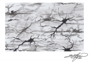
The advantage of the Golgi stain method is that it allows for the detailed study of few neurons, compared to simply staining all neurons in a given space. This makes it possible to study and visualize the structure and nuances of a single neuron.
Sarrah Hussain
Golgi Staining
India ink on paper
6”x4” -
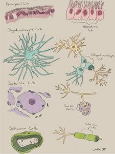
Oligodendrocytes and Schwann cells are both supporting glial cells, which serve to myelinate, and therefore insulate, neuronal axons. Oligodendrocytes are found strictly in the central nervous system (CNS), and one cell can myelinate multiple sections of an axon, as well as across multiple neurons. On the contrary, Schwann cells are found only in the peripheral nervous system (PNS), and each Schwann cell forms a singular sheath along a neuron.
Sarrah Hussain
Digital painting – Photoshop -
Microglia are a form of glial cells that serve as the “immune system” of the nervous system. In its normal resting state, microglia are small, thin, and largely non-visualized. However, upon pathological development or disease progression, microglial activation results in inflammation. Fully activated microglia appear larger, swollen, and exert neuroprotective properties. However, if chronically inflamed, overactive microglia can also display neurotoxic properties and cause unintended cell damage.
Sarrah Hussain
Watercolor on paper
8”x10” -
Diagram of the twelve cranial nerves, arising ventrally from the cerebrum and brainstem.
Sarrah Hussain
Watercolor and ink on paper
8”x10” -
This illustration shows the different cells involved in smelling with an emphasis on the olfactory ensheathing glia cells.
Viola Yu
Digital Illustration -
Illustration of the various types of glia cells.
Viola Yu
Digital Illustration -
Neurotransmitter dopamine in water, air-dried, polarized light microscopy at 100X.
Viola Yu
Gouache on Mixed Media Paper -
Stippled and line artwork showing the different pyramidal cells and how damage can move through the spinal cord as it follows the corticospinal tract.
Viola Yu
Ink on Mixed Media Paper -
This figure shows the different cranial nerves and what muscle/organs they affect.
Viola Yu
Digital Illustration -
This animation shows how one neuron can release neurotransmitters across the space between another neuron (synaptic cleft) that then in turn excites the receiving neuron.
Viola Yu
Digital Animation -
This animation demonstrates how the locomotor central pattern generator (CPGs) within the nervous system work. In the beginning of the animation it shows how even when a cat with a spinal injury when placed on a treadmill can still resume walking motions in its legs. That can all be accredited to CPGs. To explain it, we show different senarios with different cells and how they interact with one another. An endogenous burster cell (cell X) that has an excitatory synapse with cell A will cause cell X and cell A to activate at the same time and rhythm as one another (as demonstrated with tick marks along the line). When you introduce a tonically active cell (cell B) that is not synapsed with either cell X or cell A it continuously is activated. When you add an interneuron that has an inhibitory synapse to cell B, and an excitatory synapse from cell X, when you activate the endogenous burster (cell X), it causes cell A to be activated and the interneuron to become activated. With an activated interneuron it then inhibits cell B, therefore creating a pattern of when cells X is active, cell A is also active, but cell B becomes inactive since the interneuron inhibits it. When cell X is inactive, it causes cell A to become inactive and allows cell B to be active as it is not being inhibited by the interneuron. This creates a pattern of cell X and cell A being on when cell B is off and cell B is on when cell X and cell A are off. Then take for example that cell A is the injured cat’s left feet and that cell B is the cat’s right feet. That is how CPGs such as endogenous bursters can help a spinally injured cat to continue to walk when placed on a treadmill.
Viola Yu
Digital Animation -
Endorphins are neurochemicals that are produced in the brain that give you that “Runner’s high” when exercising.
Viola Yu
Digital Illustration
CIRCADIAN RHYTHMS CHAPTER:
-
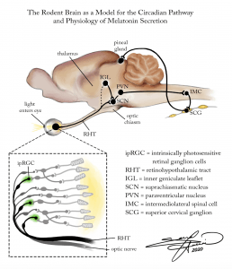
The rodent brain serves as a model for the circadian pathway and the physiology of melatonin secretion. First, light enters through the eye and is detected by special, intrinsically photosensitive retinal ganglion cells (ipRGC). This signal travels not through the optic nerve, but rather the retinohypothalamic tract (RHT), and synapses onto the suprachiasmatic nucleus (SCN), which is the internal biological clock of the body. Parallel signals also travel to the inner geniculate leaflet (IGL), which also then communicates to the SCN. The signal is then sent to the paraventricular nucleus (PVN), which then travels to intermediolateral spinal cells (IMC). These spinal cells then synapse onto superior cervical ganglion cells (SCG), which then ultimately innervate the pineal gland. From here, the pineal gland releases the hormone melatonin, which plays a key role in regulating the body’s circadian rhythm.
Sarrah Hussain
Digital Painting – Photoshop -
The Biological and Behavioral Mechanisms of Familial Advanced Sleep-Phase Syndrome:
Familial Advanced Sleep-Phase Syndrome, or FASPS, can result from a point mutation in the per2 gene, located on the 2nd chromosome. This mutation results in a disruption of healthy Per2 protein transcription, and as a result the protein is synthesized in too low quantities, as well as in a hypo-phosphorylated state. These mutated Per2 proteins result in a shortening of the circadian cycle. In other words, a person with FASPS experiences a faster or “advanced” internal biological clock, compared to the average adult. As a result, they consistently wake up much earlier (approx 2-4 hours earlier) and go to bed much earlier in the day.Sarrah Hussain
Digital Painting – Photoshop -
The Role of PER and CLOCK Genes in the Regulation of 24-Hour Circadian Cycle
Sarrah Hussain
Digital Animation – Autodesk Sketchbook
8”x10” -
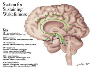
When the neuropeptide orexin is activated, it excites/activates neurons in the cortex and other regions of the brain to keep it awake.
Viola Yu
Digital Illustration -
To activate the REM cycle different parts of the brains sends different neurotransmitters that eventually sends signals across the cortex of the brain.
Viola Yu
Digital Illustration -
To keep the brain asleep, the REM cycle will activate (shown with green dots) and deactivate (shown with red dots) certain parts of the brain. Generally, the brain will inactivate, or inhibit, wake-promoting neurons. For example, the laterodorsal tegmental nuclei (SLD or LDT) produces muscle paralysis by activating the ventromedial medulla which in turn hyperpolarizes (or inactivates) the motor neurons to prevent you from acting out your dreams.
Viola Yu
Digital Illustration -
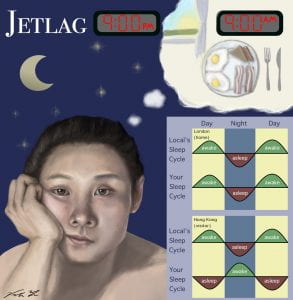
This illustrates the effects one’s sleep schedule when crossing over to different time zones quickly (such as when flying or driving). This phenomenon when your body’s circadian rhythm is out of sync from the local’s is called Jetlag. The graph on the bottom right corner demonstrates the sleep-wake cycle of the traveler compared to the local’s cycle when at home and when traveling away from home.
Viola Yu
Digital Illustration
SLEEP AND DREAMING CHAPTER:
-
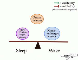
The mechanisms of narcolepsy can be explained by understanding the roles and relationships of orexin-producing neurons in the brain. Orexin, or hypocretin, is known for regulating arousal, wakefulness, and appetite. As explained visually in the animation, orexin-producing neurons act differentially on various parts of the brain to regulate the states of sleepfulness and wakefulness in a seesaw fashion. When orexin neurons provide excitatory input to monoaminergic neurons, and indirectly suppress GABA-ergic neurons, wakefulness is achieved. When orexin activity decreases, GABA-ergic neurons are more active, and also exert an inhibitory effect on orexin and monoaminergic neurons, and thus a sleep-state is induced. However, the sleep disorder of narcolepsy is characterized by inactive orexin-producing neurons. Without this regulatory neuropeptide, there is no preferential influence on either GABA-ergic or monoaminergic neurons, and so the brain is caught in an unstable alternation between sleepfulness and wakefulness.
Sarrah Hussain
Digital Animation – Autodesk Sketchbook -
A major region of the brain associated with dreaming is the amydala and hippocamus that can be found within the temporal lobe, curving up into the center of the brain.
Viola Yu
Digital Illustration
PARENTING CHAPTER:
-
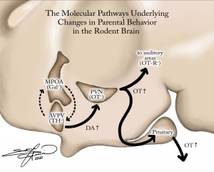
The rodent brain serves as a model for the molecular pathways underlying the changes seen in parental behavior, upon visual and tactile stimulation from offspring. Visual and tactile cues from rodent pups first stimulate the anteroventral periventricular nucleus (AVPV), which releases tyrosine hydroxylase to the medial preoptic area (MPOA). The MPOA then releases the neuropeptide galanin in a loop back to the AVPV. The AVPV also then communicates forward via increased secretion of dopamine (DA) to the paraventricular nucleus (PVN). The PVN then increases oxytocin (OT) secretion and sends signals to both the auditory areas in the upper cortex, as well as the pituitary gland. The pituitary gland then also increases OT levels and delivers this hormone throughout the endocrine system to the rest of the body, to induce social-bonding and parental behaviors.
-
Scientific experimentation found that when rat pups signaled to adult female rat the adult female rat increased maternal care and increased oxytocin release. When pup signaling occurred in the presence of an adult male rat, the adult male rat decreased their aggression and therefore became less likely to attack the rat pups.
Viola Yu
Digital Illustration

