-
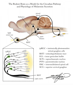
The rodent brain serves as a model for the circadian pathway and the physiology of melatonin secretion. First, light enters through the eye and is detected by special, intrinsically photosensitive retinal ganglion cells (ipRGC). This signal travels not through the optic nerve, but rather the retinohypothalamic tract (RHT), and synapses onto the suprachiasmatic nucleus (SCN), which is the internal biological clock of the body. Parallel signals also travel to the inner geniculate leaflet (IGL), which also then communicates to the SCN. The signal is then sent to the paraventricular nucleus (PVN), which then travels to intermediolateral spinal cells (IMC). These spinal cells then synapse onto superior cervical ganglion cells (SCG), which then ultimately innervate the pineal gland. From here, the pineal gland releases the hormone melatonin, which plays a key role in regulating the body’s circadian rhythm.
Sarrah Hussain
Digital Painting – Photoshop -

The Biological and Behavioral Mechanisms of Familial Advanced Sleep-Phase Syndrome:
Familial Advanced Sleep-Phase Syndrome, or FASPS, can result from a point mutation in the per2 gene, located on the 2nd chromosome. This mutation results in a disruption of healthy Per2 protein transcription, and as a result the protein is synthesized in too low quantities, as well as in a hypo-phosphorylated state. These mutated Per2 proteins result in a shortening of the circadian cycle. In other words, a person with FASPS experiences a faster or “advanced” internal biological clock, compared to the average adult. As a result, they consistently wake up much earlier (approx 2-4 hours earlier) and go to bed much earlier in the day.Sarrah Hussain
Digital Painting – Photoshop -

The Role of PER and CLOCK Genes in the Regulation of 24-Hour Circadian Cycle
Sarrah Hussain
Digital Animation – Autodesk Sketchbook
8”x10” -
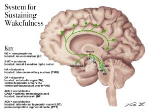
When the neuropeptide orexin is activated, it excites/activates neurons in the cortex and other regions of the brain to keep it awake.
Viola Yu
Digital Illustration -

To activate the REM cycle different parts of the brains sends different neurotransmitters that eventually sends signals across the cortex of the brain.
Viola Yu
Digital Illustration -

To keep the brain asleep, the REM cycle will activate (shown with green dots) and deactivate (shown with red dots) certain parts of the brain. Generally, the brain will inactivate, or inhibit, wake-promoting neurons. For example, the laterodorsal tegmental nuclei (SLD or LDT) produces muscle paralysis by activating the ventromedial medulla which in turn hyperpolarizes (or inactivates) the motor neurons to prevent you from acting out your dreams.
Viola Yu
Digital Illustration -

This illustrates the effects one’s sleep schedule when crossing over to different time zones quickly (such as when flying or driving). This phenomenon when your body’s circadian rhythm is out of sync from the local’s is called Jetlag. The graph on the bottom right corner demonstrates the sleep-wake cycle of the traveler compared to the local’s cycle when at home and when traveling away from home.
Viola Yu
Digital Illustration


