-

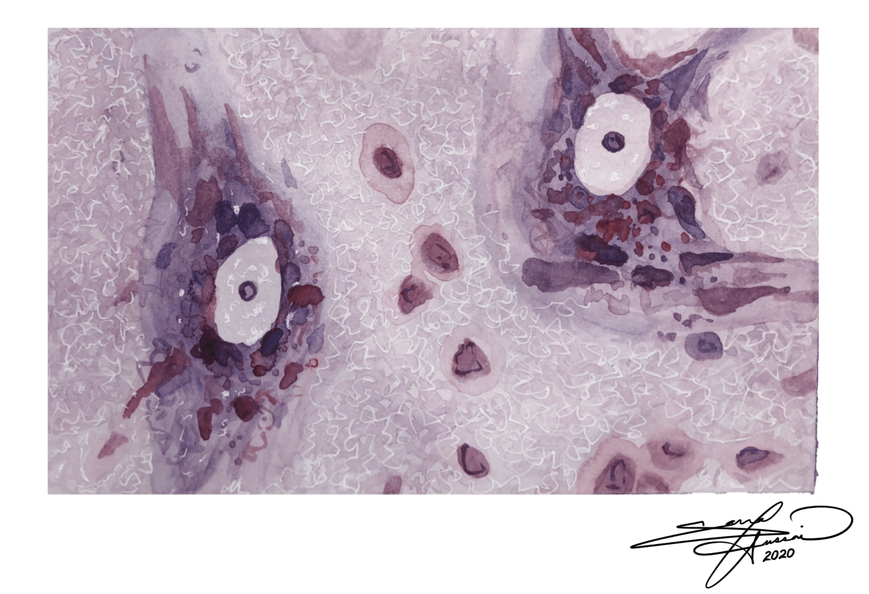
Using a Cresyl Violet stain would be best for evaluating the sex differences in the number of neurons in the brain, because this stain highlights all neurons and cells in a given area. This makes it possible to count and compare number of neurons in the brain between sexes.
Sarrah Hussain
Cresyl Staining
Mixed media on paper
6”x4” -
The advantage of the Golgi stain method is that it allows for the detailed study of few neurons, compared to simply staining all neurons in a given space. This makes it possible to study and visualize the structure and nuances of a single neuron.
Sarrah Hussain
Golgi Staining
India ink on paper
6”x4” -

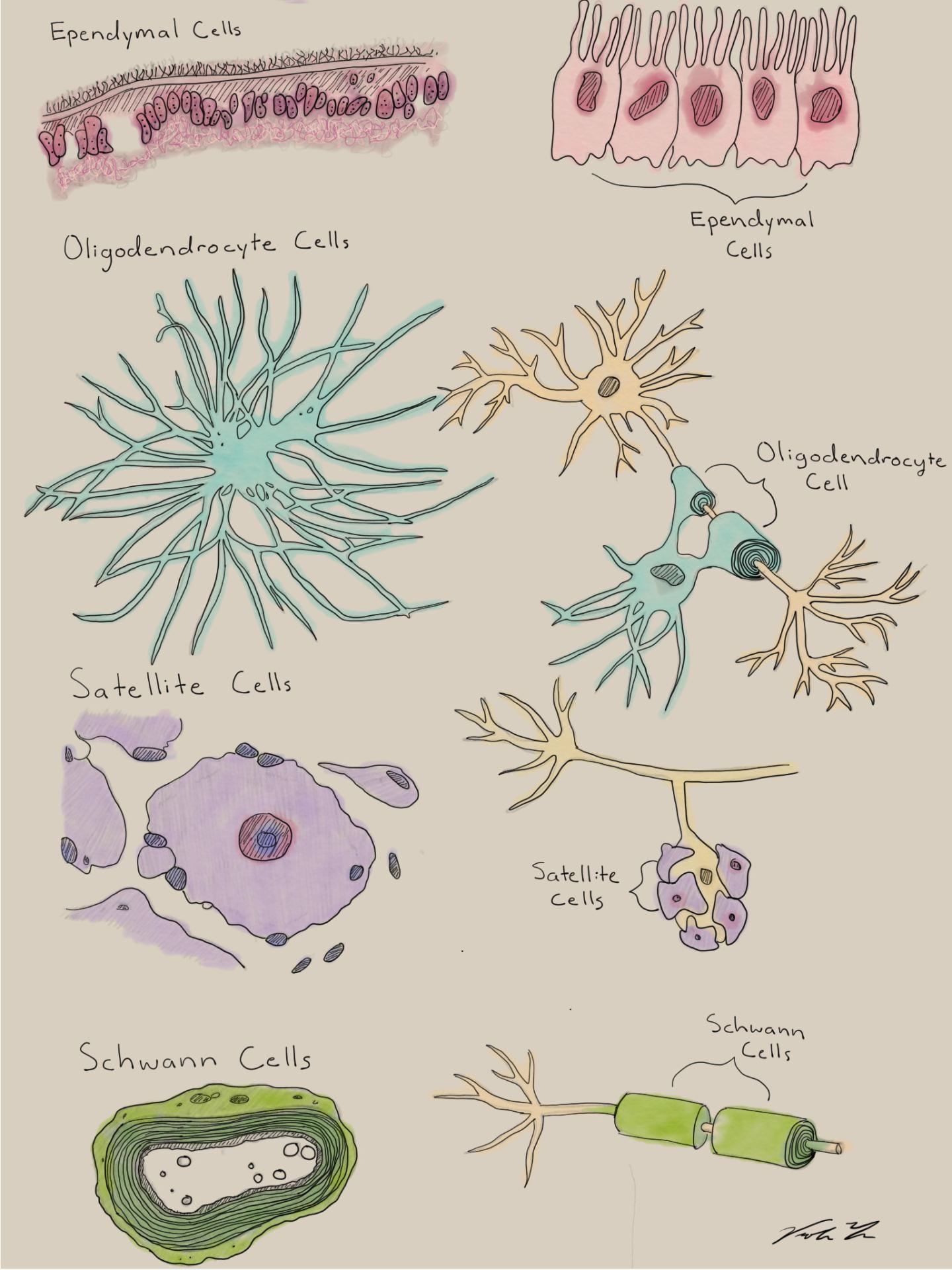
Oligodendrocytes and Schwann cells are both supporting glial cells, which serve to myelinate, and therefore insulate, neuronal axons. Oligodendrocytes are found strictly in the central nervous system (CNS), and one cell can myelinate multiple sections of an axon, as well as across multiple neurons. On the contrary, Schwann cells are found only in the peripheral nervous system (PNS), and each Schwann cell forms a singular sheath along a neuron.
Sarrah Hussain
Digital painting – Photoshop -

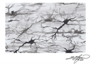
Microglia are a form of glial cells that serve as the “immune system” of the nervous system. In its normal resting state, microglia are small, thin, and largely non-visualized. However, upon pathological development or disease progression, microglial activation results in inflammation. Fully activated microglia appear larger, swollen, and exert neuroprotective properties. However, if chronically inflamed, overactive microglia can also display neurotoxic properties and cause unintended cell damage.
Sarrah Hussain
Watercolor on paper
8”x10” -

Diagram of the twelve cranial nerves, arising ventrally from the cerebrum and brainstem.
Sarrah Hussain
Watercolor and ink on paper
8”x10” -

This illustration shows the different cells involved in smelling with an emphasis on the olfactory ensheathing glia cells.
Viola Yu
Digital Illustration -
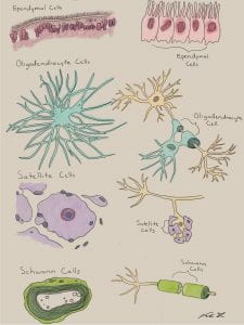
Illustration of the various types of glia cells.
Viola Yu
Digital Illustration -
Neurotransmitter dopamine in water, air-dried, polarized light microscopy at 100X.
Viola Yu
Gouache on Mixed Media Paper -

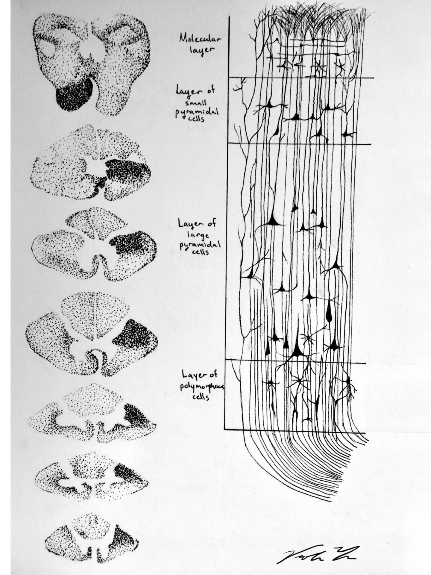
Stippled and line artwork showing the different pyramidal cells and how damage can move through the spinal cord as it follows the corticospinal tract.
Viola Yu
Ink on Mixed Media Paper -

This figure shows the different cranial nerves and what muscle/organs they affect.
Viola Yu
Digital Illustration -

Endorphins are neurochemicals that are produced in the brain that give you that “Runner’s high” when exercising.
Viola Yu
Digital Illustration