Introduction.
Much of this post is an overview of comparative vertebrate neuroanatomy. The major structures of the vertebrate brain as well as their primary function will be presented (focusing on their relevance to humans). At the end, I will show you how mother nature got from a fish brain to a human brain during the course of human evolution.
I took a comparative vertebrate anatomy course from Clark Hubbs, a well-known zoologist, at the University of Texas when I was an undergraduate about 100 years ago (actually only 55 years ago, but seems like 100 😀). This course turned out to be very influential in my professional development. In fact, this post draws heavily on what I learned many years ago. By comparing the organ systems of different vertebrates, Hubbs explained how they changed during evolution. Being a psychology major, I was most interested in the brain.
Perhaps the most profound take-home lesson for me was that while the anterior 20% of the vertebrate brain (which includes the cortex) changed dramatically during human evolution, the posterior 80% (the brainstem) changed remarkably little. If you want to understand the complex workings of the human cortex and consciousness, you pretty much have to study humans or our close primate relatives. However, you can learn a lot about the human brainstem by studying any vertebrate. There is definitely a vertebrate brainstem plan, with different parts doing pretty much the same sorts of things in different vertebrates.
In evolutionary parlance, we would say that the brainstem is evolutionarily conserved. Conserved traits are those that are designed so well by Mother Nature that they do not change much over evolutionary time. There have, of course, been brainstem changes in human evolution, often related to the human cortex acquiring functions controlled exclusively by the brainstem of lower vertebrates. However to me, the vertebrate similarities are more remarkable than the differences.
The Primitive Vertebrate Brain Plan.
The first vertebrates were fish. The primitive vertebrate brain seen in Figure 1 is basically a schematic of a fish brain. While an actual fish brain wouldn’t look exactly like my schematic, you would be able to see the similarities. It should also be mentioned that the brain is a bilateral organ with every structure occurring in both the right and left sides of the brain.

Let’s get some terminology out of the way first. There are several schemes for dividing up the brain (see figure 1). The simplest is to divide it into 3 parts: forebrain, midbrain and hindbrain. However, anatomists like to break it down further and use more technical terms. A more technical term for forebrain is prosencephalon. The prosencephalon can be further subdivided into the telencephalon and diencephalon. The midbrain becomes the mesencephalon while the hindbrain becomes the rhombencephalon. The rhombencephalon can be further subdivided into the metencephalon and mylencephalon.
When considering the brains of nonhuman vertebrates, anterior refers to the front part of the brain, posterior to the back, dorsal to the top, and ventral to the bottom. Because the human brain has a different orientation in relation to the body, human neuroanatomists use the terms superior and inferior to replace dorsal and ventral when referring to the brain.
You also need to know that brain tissue consists of either grey matter or white matter. Grey matter is composed mainly of neuron cell bodies (whose membranes are grey), while white matter comes from myelinated fiber tracts that interconnect different areas of grey matter (axonal membranes in these fiber tracts are covered by white myelin sheaths providing the fiber tracts white color). Throughout the brain there are clusters of grey matter interspersed among the white matter. These clusters of neuron cell bodies are called nuclei by neuroanatomists (which is different from how this term is used by a cell biologist) and there are many different nuclei, and collections of nuclei, throughout the brain.
And finally you need to know that the brain is a massive parallel processor with different types of processing able to occur simultaneously. This division of labor is accomplished by having different nuclei and neural circuitries for different functions. These circuitries often do not interact until their modular processing is complete.
Telencephalon.
The olfactory bulb in the front of the brain (figure 2) receives input from the olfactory nerve and provides the initial unconscious processing for the sense of smell. The olfactory nerve is the most anterior of the 12 pairs of cranial nerves that enter the brain. Cranial nerves are composed of both sensory and motor neurons and service mainly the head and face. The rest of the body is similarly serviced by 31 pairs of spinal nerves that connect with the spinal cord. However, one cranial nerve, the vagus nerve, services both parts of the head and as well as many organs in the lower body (vagus means “wandering”).
While most vertebrates have relatively large olfactory bulbs, humans have small ones. This difference reflects olfaction (and pheromones) playing a much larger role in the social/sexual lives of most non-human vertebrates, while vision subserves much of this function in humans. In fact, a general principle is that the relative size of a brain structure is related to its importance for a particular species. Not only is the olfactory bulb relatively small in humans, there are also fewer types and numbers of olfactory receptors. In addition, a secondary vertebrate olfactory system, the vomeronasal system, is either vestigial or absent in humans. As Hubbs put it, “most vertebrates smell in color, while humans smell in black and white.”

The olfactory system is thought by some to be the earliest and most primitive sensory system and plays by different anatomical rules than other sensory systems. In humans, the pathway from the olfactory bulb to the cortex is different than for the other senses, and more processing occurs below the level of conscious awareness.
The dorsal bump in the telencephalon, and everything inside of it, is called the cerebrum (see figure 2). The cerebrum has a thin outer covering of grey matter called the cerebral cortex, or just cortex. The cortex is essentially a massive nucleus. During human evolution, the size of cortex expanded dramatically and acquired some of the sensory and motor capabilities managed by the brainstem of lower vertebrates. In addition, this evolution eventually led to the complexities of human consciousness. More about the cortex and consciousness in another post.
Much of the inside of the cerebrum is white matter consisting of fiber tracts interconnecting different parts of the cortex to each other as well as to nuclei throughout the brain. Most connections are “2-way streets.” Buried in the cerebrum’s white matter are two important systems, the limbic system and the basal ganglia. Both consist of a number of interconnected nuclei. Although both systems operate below the level of conscious awareness, the outcomes of their processing play major roles in assisting the conscious activities of the cortex.
The nuclei of the limbic system include the hippocampus, septum, amygdala, and nucleus accumbens. The hippocampus and septum are necessary to help the cortex consolidate short-term memories (transient electrical events) into long-term memory (relatively permanent structural changes). In fact, the memory problems of Alzheimer’s Disease begin with a loss of acetylcholine neurons that provide input into the hippocampus which, in turn, impairs the hippocampus’s ability to support the cortex’s consolidation process. In the early stages of Alzheimers, the old long-term memories are still there, but the person has difficulty forming new ones. As a result, such an Alzheimer’s patient can regale you with stories of their youth, but can’t remember what happened to them yesterday. As the disease progresses the memory problems worsen as both the hippocampus and cortex begin to degenerate and existing long-term memories are eventually lost as well.
The amygdala, on the other hand, interprets emotional situations to provide input important in forming emotional memories in the cortex. Post-traumatic stress disorder (PTSD) is thought to arise from intensely traumatic events causing amygdala hyperreactivity. The resulting memory can later be reactivated by any situation remotely reminding the person of the traumatic event, causing an incapacitating “fight or flight” reaction.
And finally, the limbic system’s nucleus accumbens helps the cortex to interpret the hedonic value of all forms of reward (i.e. food, drink, sex, warmth, etc) and also plays a central role in providing the motivation necessary for both learning and drug addiction (many experts consider drug addiction to be a type of maladaptive learning).
The different nuclei of the basal ganglia are critical for helping the cortex plan and execute motor movements. The globus pallidus is more important in initiating movement, while the caudate nucleus/putamen, in terminating it. Proper functioning requires dopamine input into the caudate nucleus/putamen from a midbrain nucleus called the substantia nigra. Parkinson’s Disease arises from the selective degeneration of the substantia nigra, which removes this dopamine input.
Huntington’s Disease is another motor disease of the basal ganglia. However, in this case, a loss of GABA-secreting neurons in the basal ganglia itself is the cause. (GABA is the principle inhibitory neurotransmitter throughout the brain.) As the disease progresses other types of neurons in the basal ganglia and cortex are also affected. Both motor diseases are sometimes accompanied by psychotic symptoms suggesting that the basal ganglia, through its connections with the cortex, may play a role in psychological functioning.
Diencephalon.
The diencephalon sits right behind the telencephalon and contains the thalamus, epithalamus (containing the pineal gland), and hypothalamus. Each of these general areas contain multiple nuclei with different functions. The classification scheme presented here considers the diencephalon as the beginning of the brainstem (although some neuroanatomists consider the mesencephalon to be the beginning. It can be confusing that different neuroanatomists don’t always agree on terminology 🤬). The pituitary gland sits just below the hypothalamus and just anterior to the pituitary is a distinctive landmark called the optic chiasm. The optic chiasm is where the optic nerve enters the brain and is caused by half of the nerve fibers on each side crossing over to the other side.
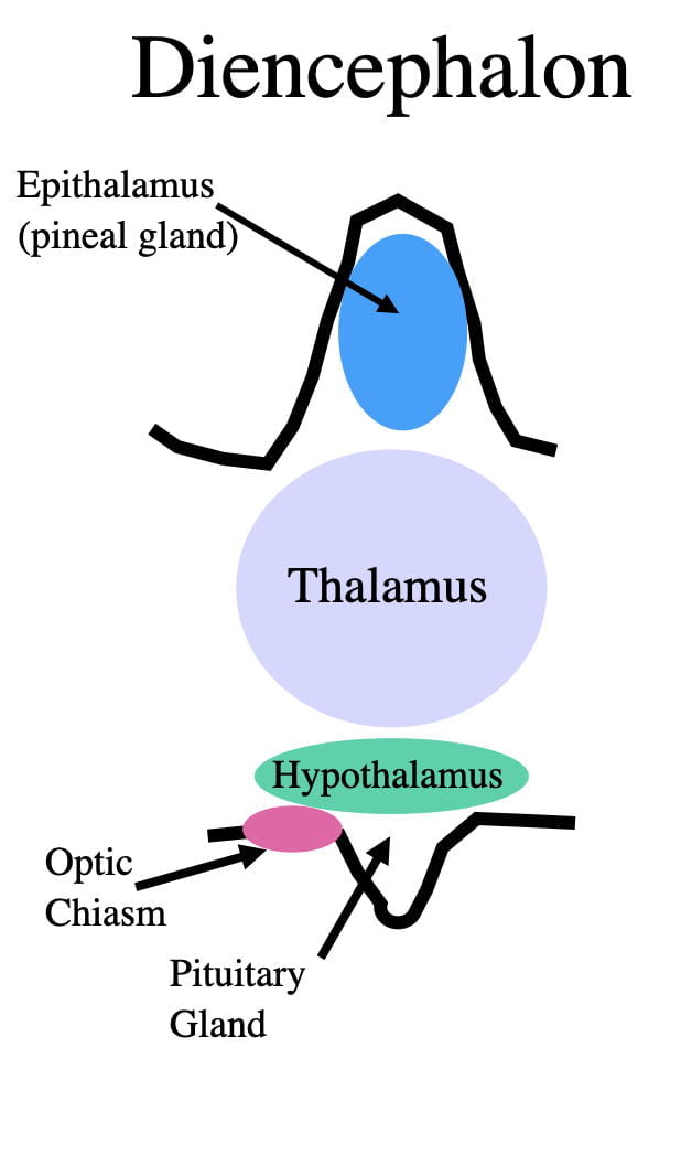
Moving to the top of the diencephalon, the epithalamus contains the pineal gland. Earlier I stated that all structures in the brain are bilaterally represented. I lied! The pineal gland is the human brain’s only unilateral structure with the 2 halves fused into a single midline structure. This unique arrangement led the famous scientist/philosopher Descartes to suggest that the pineal gland is the “seat of the soul.” I’ll leave it to others to debate that suggestion.
In vertebrates, every cell in the body has its own circadian clock whose time keeping is linked to the daily synthesis and degradation of certain proteins. Circadian clocks optimize physiological and behavioral adaptation to different times of the day. For example, if food is available only at certain times of the day, biological clocks prepare the organism to physiologically and behaviorally take advantage. Virtually all physiological processes show circadian rhythms, functioning at their highest level at certain times of the day or night.
Proper circadian timing is clearly important for humans. The tiredness and malaise from jet lag is because the circadian clocks are temporarily decoupled from the local time. Fortunately recovery typically takes only a few days and does not usually have lasting effects. However, chronic jet lag, as seen in shift workers, is associated with significantly increased illness, disease, and mortality.
A problem with each cell in the body having its own circadian clock is that each clock’s periodicity often differs slightly from 24 hours, and also slightly from each other. Allowed to “free run”, the cellular clocks would quickly get out of sync with the 24-hour cycle and with each other. This asynchrony would cause the dysfunctions of jet lag. Mother nature has solved the problem by having one clock to rule them all. This clock is known as the “master clock”, which keeps all the other clocks synchronized with both local time and with each other.
However, the master clock may also have a periodicity that isn’t exactly 24 hours. For example, in humans, the master clock, for unknown reasons, has an average periodicity of around 25 hours (it can vary a bit from one individual to the next). The master clock solves this problem by “re-setting itself” to the local time every day. Resetting is triggered most often by light onset in the morning although other sensory cues that occur at approximately the same time every day can also be used (such as the crowing of a rooster, or the ringing of an alarm clock). By resetting the human master clock back one hour each day, the body’s time keeping is kept in step with that of the environment. However, the human master clock is capable of retarding or advancing itself only a couple hours each day. So if your clock gets more than a couple of hours out of sync with the local time you experience jet lag until the master clock can resynchronize itself with the local time.
The pineal gland is the master clock of reptiles and birds. In fact, in some birds, where light can penetrate their paper-thin skulls, light-sensitive photopigments similar to those in our eyes allow the pineal to actually sense light to reset the master clock. Even stranger, in some lizards the pineal extends through the skull to become a third eye in the forehead. However, this light-sensing “eye” is not for vision but for entraining the master clock.
The pineal gland of vertebrates, including humans, also exhibits a circadian rhythm in the secretion of the hormone, melatonin. Melatonin concentrations are highest at night while sleeping and promote sound sleep. In fact, melatonin is sold over-the-counter in health food stores and pharmacies for this purpose. Also supporting melatonin’s sleep-promoting effect is that the restless sleep from watching tv or using a phone or tablet just before bed-time, is because the blue light from these devices inhibits nighttime melatonin secretion. in addition, after flying on an airplane to distant locations, melatonin, taken just before bedtime not only promotes sleep, it also shortens the period of jet lag.
In temperate-climate vertebrates, melatonin has also been implicated in regulating seasonal behaviors such as hibernation, migration, food hoarding, etc. These seasonal changes, in some cases, are influenced by the increased melatonin secretion during the longer nights of the winter. There is some evidence for human seasonality (e.g. seasonal affective disorder and births peaking in summer and fall), but the data are not as robust as for many other vertebrates. Perhaps that’s because much of human evolution occurred in the tropics where the seasonal changes are not as great.
The structure immediately below the epithalamus/pineal gland is the thalamus. The job of the thalamus is to serve as a relay station to send sensory information up to the cortex. With the exception of olfaction, sensory information entering the brain and spinal cord makes its way to the thalamus. Neurons in the thalamus then relay this sensory information up to the cortex. To accomplish this task, the thalamus contains a number of different nuclei, each specializing in relaying a different type of sensory information. Vision, hearing, touch, taste, temperature, pain etc. are then relayed separately to different cortical areas for conscious processing and awareness.
The structure below the thalamus is appropriately called the hypothalamus. While relatively small (in humans about the size of an almond), the hypothalamus plays an oversized role in physiology and behavior. In terms of behavior, the hypothalamus controls the primitive, largely unconscious,”animalistic” behaviors that we share with other vertebrates. Hubbs, my comparative anatomy teacher, had a great mnemonic for remembering the names of these behaviors. He called them the “4 F’s: feeding, fighting, fleeing, and mating.”
When I was an undergraduate 55 years ago, we referred to the pituitary gland, just below the hypothalamus, as the master gland because it produces a large number of different hormones that, in turn, control the other endocrine glands of the body. However, we now know that the hypothalamus, through hormonal and neural connections, controls the pituitary. So, the hypothalamus is the true master gland playing a critical role in proper body functioning and homeostasis.
In mammals, the hypothalamus also has a third important function. Unlike reptiles and birds, a nucleus in the hypothalamus, the suprachiasmatic nucleus, serves as the master clock. (Suprachiasmatic refers to its location immediately above the optic chiasm.) This nucleus makes sure all the mammalian circadian clocks are synchronized to the time of day and to each other.
Why the mammalian master clock would be different from other vertebrates likely has to do with an evolutionary change in the relative position of the pineal gland (which I’ll explain in more detail later). Instead of being on the dorsal surface of the brain, as it is in birds and reptiles, the mammalian pineal is buried deep inside the brain, presumably impairing its ability to entrain its daily time keeping. A better option for mammals is to use light detected by the eye rather than the pineal to reset the clock. The suprachiasmatic nucleus, which is just above the optic chiasm, is well situated for this purpose. In fact a small number of optic nerve fibers synapse in the suprachiasmatic nucleus, making it highly efficient for light to reset the mammalian master clock each day.
Mesencephalon (Midbrain).
The mesencephalon or midbrain consists of a dorsal tectum and a ventral tegmentum. The tectum contains the primary sensory and motor centers of all non-mammalian vertebrates. As mentioned earlier, these functions have been acquired by the cortex in mammals. The tegmentum, like other parts of the ventral brainstem, contains many fiber tracts running up and down the brain. There are also 2 cranial nerves that connect with the brain here. In addition, there is the most anterior part of a diffuse polysynaptic pathway running up the brainstem called the reticular activating system or just reticular system.
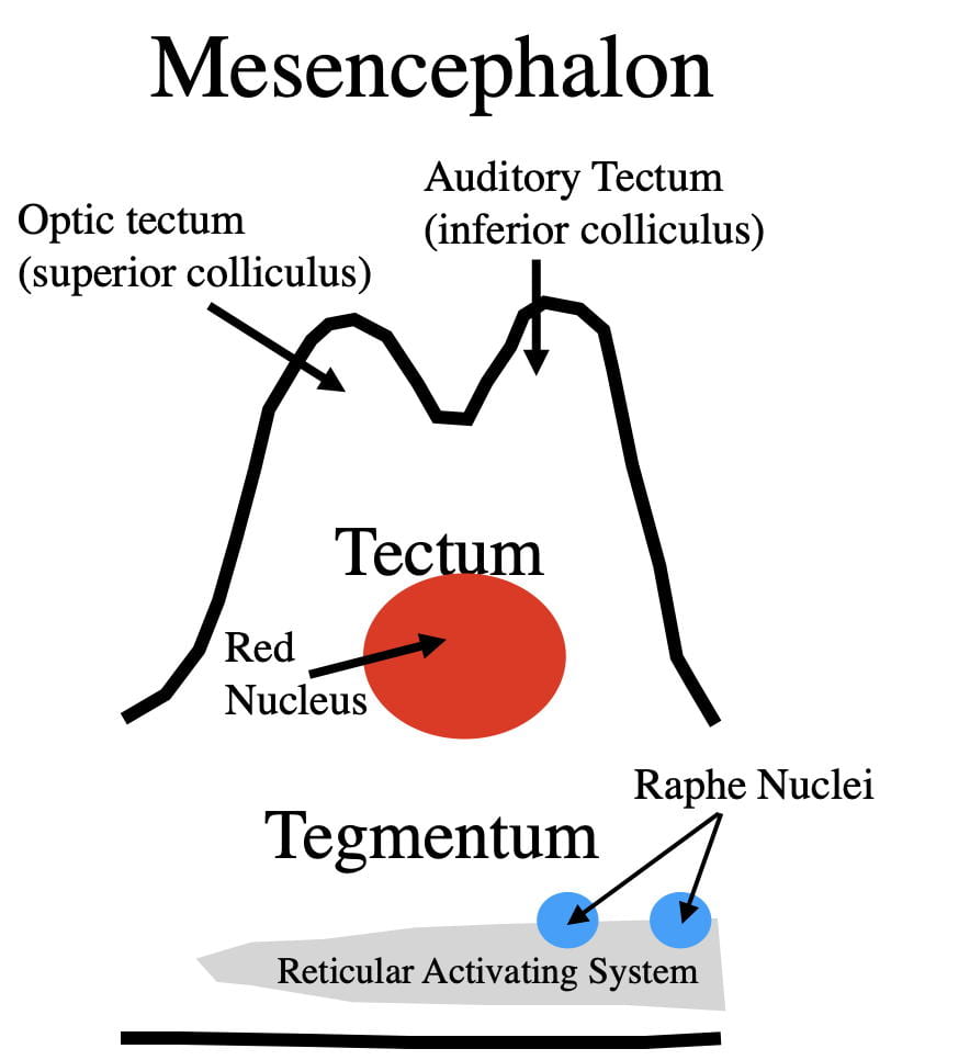
Within the tectum, sensory function is dorsal to motor function. The first “minibump” of the tectum is the optic tectum, the primary visual processing center of all nonmammalian vertebrates. In mammals the cortex has acquired these visual processing functions to more effectively bring them under conscious control. Because of its primary role, the optic tectum is relatively larger in non-mammalian vertebrates. (In humans, the optic tectum is called the superior colliculus)
There are pluses and minuses to having visual processing in the tectum. The advantage is the ability to respond quickly to visual input. The slowest aspect of neural communication is synaptic neurotransmission. Any response managed by tectal processing requires fewer synapses and is inherently quicker than one managed by the cortex. The advantage to cortical processing, on the other hand, is that the processing is brought under greater conscious control. Conscious processing allows for more complicated and nuanced responses that can be tailored to a wider variety of circumstances. In the case of mammals, and particularly humans, a larger response repertoire is more adaptive than responding quickly. However, cortical processing remains quick enough for most tasks (we’re talking fractions of a second difference).
We humans still have a functional optic tectum/superior colliculus that is still processing visual input. So what is it doing in humans? There are some visual reflexes, even in mammals, that must be exceedingly quick to be effective. To this end, the superior colliculus controls eye and head movements that allow us to quickly focus our gaze on objects that draw our attention. These responses occur automatically and do not require conscious processing. If the cortex were to control these responses, they would be slower and less adaptive.
There is also evidence that the human superior colliculus may be capable of more complicated unconscious reflexes. For example there are retinotopic maps in the superior colliculus where locations in the retina correspond to specific locations in the superior colliculus. This relationship suggests that the superior colliculus, like the cortex, can create a representation of the visual field. Perhaps this ability underlies a very interesting phenomenon called “blindsight.” This rare phenomena is seen in individuals blinded by damage to their visual cortex while leaving the tectum undamaged. Very strangely, such blind individuals have been observed to respond appropriately to certain visual stimuli. However, if you ask them why, they are not consciously aware of the reason. Such quick, unconscious, but complicated reflexes could be highly adaptive in responding to dangerous (or rewarding) stimuli that require an absolutely immediate response to be effective.
Cortical versus tectal visual processing illustrates another interesting evolutionary principle. When mother nature figures out a “better way” of doing something, she often doesn’t get rid of the old way. She simply superimposes the new way on top of the old way. This approach often creates some redundancy, illustrating yet another important evolutionary principle. The more important a brain process is to fitness and survival, the more likely it is to be controlled by redundant backup systems. That way if one system fails, another has some potential to step in and take over.
The evolution of mammalian hearing is similar to that of vision. The auditory tectum is the primary auditory center for all non-mammalian vertebrates. However in mammals, the cortex has acquired this function. However, the human auditory tectum/inferior colliculus continues to mediate quick unconscious reflexes such as the auditory startle reflex.
In the ventral tectum just below the sensory areas is the Red Nucleus, the primary motor center of the primitive vertebrate. This nucleus allows sensory information processed in the tectum to be reflected out to the muscles for quick, but unconscious, motor responses. There are 2 multisynaptic pathways from the Red Nucleus to the ⍺-motoneurons of the brain and spinal cord that are collectively called the extrapyramidal motor system. The ⍺-motoneurons then carry the message from the brain and spinal cord to the muscles to cause muscle contractions. The extrapyramidal system can produce unconscious, reflex-like behaviors when acting upon sensory information from the tectum. However, in mammals the extrapyramidal system can also be accessed by the cortex allowing for conscious body movements as well.
If you guessed that there must also be a pyramidal motor system, you’d be correct! The pyramidal system is an evolutionarily newer motor pathway allowing the mammalian motor cortex another way to access ⍺-motoneurons. In fact, its pathway is even more direct than the extrapyramidal motor system making it look like it should be the primary motor system. However, it is actually a supplementary motor system that adds additional motor functionality to mammals. (As an aside, the pyramidal motor system was named by an over-imaginative neuroanatomist who thought this pathway, running along the base of the brain, looked like a pyramid in cross section.).
The primary purpose of the pyramidal motor system is to allow the cortex to control conscious dextrous movements of the distal limbs. A good example would threading a needle. To do so requires feedback from the sense of touch that must then be quickly reflected out to the appropriate finger muscles to be effective. The relative size of the pyramidal motor system in mammals correlates with the degree of manual dexterity. Dogs have relatively small systems while humans have large ones. The pyramidal motor system provides humans with the highest manual dexterity of any species on the planet. However, if you’re guessing that humans have the most developed pyramidal motor system, you’d be wrong. It turns out that many monkeys have even more developed systems. That’s because monkeys have dexterity in both their hands and their feet.
However, the two systems typically work together in humans in a complementary fashion. The extrapyramidal system is better at controlling the muscles of the truck and proximal limbs, while the pyramidal system is particularly good at controlling the movements of the hands and feet. There is some overlap, allowing one system some capability to take over if the other doesn’t function properly. For example, monkeys with nonfunctional pyramidal systems are clumsy at dextrous hand and foot movements but are otherwise able to do many things relatively normally. On the other hand, a totally dysfunctional extrapyramidal system would likely be fatal.
In summary, the extrapyramidal system is the basic, original motor system which, in a fish, operates below the level of conscious awareness. However over evolutionary time, the extrapyramidal system was brought under conscious control while at the same time continuing to operate unconsciously as well. As mammals developed dexterity in the distal end of their limbs, the pyramidal system evolved to support this dexterity.
The tegmentum is the ventral portion of the mesencephalon. The tegmentum contains the anterior end of a diffuse, polysynaptic pathway called the reticular activating system. This system consists of 20+ interconnected nuclei, extending throughout the length of the brainstem. The reticular system receives input from all the different sensory systems and at one time was thought to be a redundant system for processing sensory input. We now know this is not the case. The reticular activating system serves mainly to influence the cortex’s state of consciousness during wakefulness and sleep. More about reticular system functioning in another post covering the neuroanatomy of sleep.
Located in the dorsal reticular system are a series of 8 Raphé nuclei. The first 2 are in the mesencephalon, 3 more in the pons, and the final 3 in the medulla. These nuclei are the brain’s source of the neurotransmitter serotonin. Despite constituting an exceedingly small fraction of the brain, the axons of the Raphé nuclei release their serotonin virtually everywhere in the brain and spinal cord. The anterior Raphé nuclei service the more anterior parts of the brain, while the more posterior nuclei service the more posterior parts of the brain and spinal cord. Serotonin is released both synaptically and extrasynaptically by the Raphé neurons to modulate overall brain functioning. (Extrasynaptic neurotransmitter release is discussed in another post in the section entitled “The Brain’s Serotonin System.)
Serotonin concentrations in the brain’s extracellular fluid show a pronounced circadian rhythm, highest when awake and active, lower when awake and inactive, even lower during non-REM sleep, and lowest during REM sleep. In fact body movement is one factor known to promote serotonin release. Brain serotonin concentrations affect such diverse phenomena as mood, sleep and sex drive.
However, serotonin is probably best known for its role in Major Depression. Selective serotonin reuptake inhibitors (SSRIs), are the first-line antidepressant drugs and clear depression by boosting brain serotonin concentrations. Behavioral methods that raise brain serotonin concentrations also relieve depression in some cases. For example, daily exercise is the best non-drug treatment for depression. And for initially clearing a depression, sleep deprivation, or selective REM-sleep deprivation, which reduces time spent in periods of low serotonin concentration, will sometimes help. However, SSRIs and behavioral methods don’t help all depressed patients and there are clearly factors other than just serotonin at work in depression. A more in depth treatment of this topic is found in another post
It is worth mentioning that serotonin is not produced exclusively by neurons in the brain. In fact, 98% of the body’s serotonin is produced outside the brain by specialized cells in the digestive tract where it acts as a hormone to influence gastric motility. However, since serotonin does not cross the blood brain barrier, the brain is directly affected only by its own serotonin.
Metencephalon.
The dorsal metencephalon is called the cerebellum, while the ventral metencephalon is the pons. The cerebellum resembles the cerebrum in possessing an outer cortex of grey matter with inner white matter interspersed with nuclei. The cerebellum is best known for its role in movement and coordination.

The cerebellum plays an important role in helping both cortical and midbrain motor areas carry out complicated motor movements. To do so, the cerebellum encodes memories of complex coordinated motor movements. These memories are sometimes referred to as “motor melodies” because the precise timing of muscle contractions underlying these movements resembles that of the musical notes necessary to produce a melody. The motor melodies of the cerebellum can be incorporated into both conscious and unconscious motor movements.
The workings of the cerebellum underly the ability of athletes, dancers, musicians, artists, and others to perform their amazingly coordinated movements. The cerebellum acquires and refines its motor melodies through experience and learning….practice, practice, practice.
As with other processes in the brainstem, the motor-melody memories are encoded below the level of conscious awareness. For example, while a person consciously knows whether or not they can ride a bicycle, they do not consciously know which muscles must contract, for how long, and in what order. That’s the job of the unconscious cerebellum. Which brings up the interesting dichotomy between being a good performer of a movement and teaching that activity to someone else. In order to perform the movement you must have the motor melodies encoded in your cerebellum, but to be a good teacher or coach, you must have the relevant (but different) information stored in your conscious cortex. That’s why a good athlete isn’t necessarily a good coach and vice versa.
The pons resembles other parts of the ventral brainstem with fiber tracts running up and down the brain, part of the reticular activating system, as well as 4 cranial nerves that enter and exit here. There are also various fiber tracts coursing through here as well. However, a nucleus worth describing in more detail is the Locus Coeruleus (See figure 5).
The neurons of the Locus Coeruleus use norepinephrine as their neurotransmitter. Like the Raphé nuclei, the Locus Coeruleus is part of the dorsal reticular system. In fact, the Raphé nuclei and the Locus Coeruleus have a lot in common. The neurotransmitters of both are classified as monoamines. Both send out unmyelinated axons to synapse both synaptically and extrasynaptically throughout the CNS. Both spontaneously release their neurotransmitters in a circadian fashion, being highest during the day and lowest during sleep. In addition, both neurotransmitters are also produced outside the brain by endocrine tissue where they act as hormones rather than neurotransmitters. Neither molecule crosses the blood brain barrier so the brain is not directly affected by concentrations outside the brain. And finally, low brain levels of both have been implicated in depression. While SSRI’s are the first-line antidepressants, selective norepinephrine reuptake inhibitors (SNRI’s), are also effective in some patients by selectively boosting brain norepinephrine levels. The role of these monoamines in depression is covered in more detail elsewhere.
However the main function of the brain’s norepinephrine is to serve as the neurotransmitter of the sympathetic branch of the autonomic nervous system. The autonomic nervous system controls the unconscious automatic processes necessary to keep brain and body functioning optimally and is composed of sympathetic and parasympathetic branches that generally have opposite effects. Activation of the sympathetic nervous system causes what are known as “fight or flight” responses to help vertebrates cope with emotion-provoking situations.
For example, in the cortex, high norepinephrine concentrations cause “vigilance,” extreme attentiveness to sensory input. Outside the brain, norepinephrine is also the neurotransmitter of sympathetic nervous system neurons that prepare the muscles and circulatory action for action. In addition, norepinephrine causes all sensory organs to operate at peak efficiency. Norepinephrine also puts the body into a catabolic state optimizing the release of stored carbohydrates and fats to supply the energy needed to deal with the emotional situation.
However, that’s not all folks. The sympathetic nervous system also activates an endocrine gland, the adrenal medulla to release norepinephrine and epinephrine into the blood where they act as hormones. Both hormones then circulate throughout the body via the blood and add to the norepinephrine being released by the sympathetic neurons. So norepinephrine is both the neurotransmitter, and hormone, of sympathetic arousal. (Epinephrine and norepinephrine are also known as adrenalin and noradrenalin respectively). Another good example of a critical biological process exhibiting redundancy!
By the way, as a teacher, I always found redundancy to be an important teaching tool. My teaching philosophy was if you throw enough cow paddies against the wall (we actually used a different term for cow paddies), eventually some of them start to stick! (A little Texas humor to break up the seriousness! 😀).
The parasympathetic system is controlled by the medulla and its role will be described in more detail in the next section.
Myelencephalon.
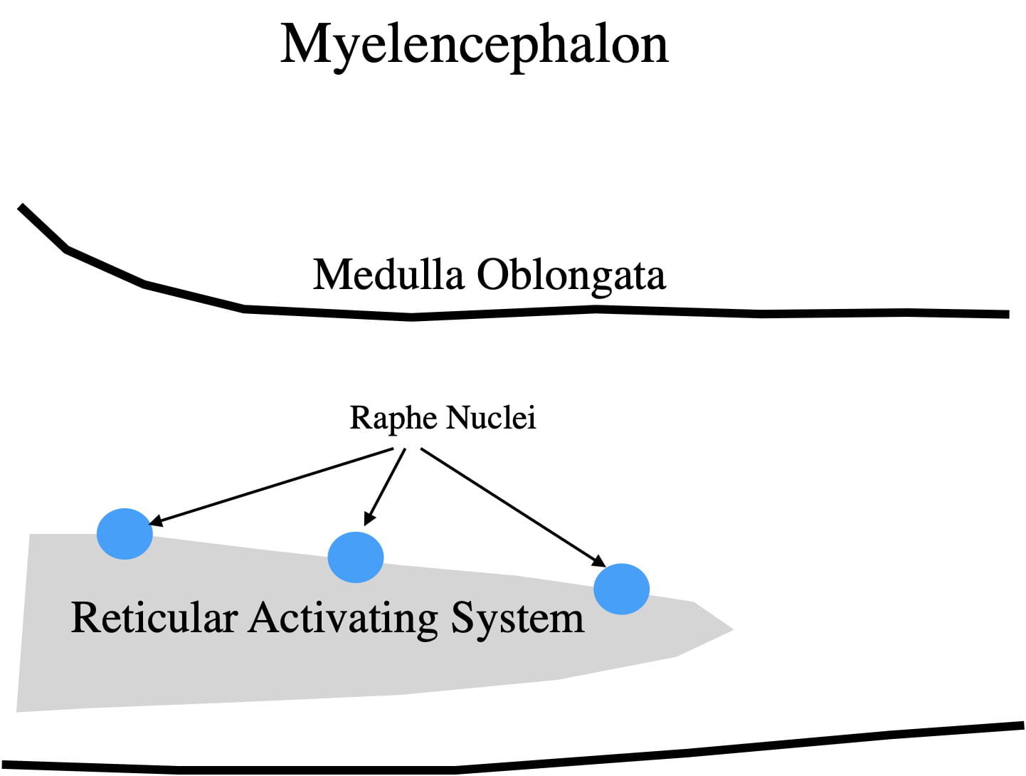
The myelencephalon contains the medulla oblongata or just medulla, the most posterior and primitive section of the brain, which connects the brain with the spinal cord. The medulla houses both sensory and motor fiber tracts from the spinal cord connecting with more anterior nuclei. The medulla also contains the most posterior part of the reticular activating system important in sleep and arousal. Also 4 of the 12 cranial nerves enter and exit the brain at this level. Again the primary purpose of the cranial nerves is to service the sensory and motor functions of the face and head. And some nuclei in this area are critical components of the parasympathetic nervous system.
The parasympathetic branch of the autonomic nervous system predominates when you are not facing an emotion-provoking situation, which for most of us, is most of the time. The parasympathetic nervous system activates the anabolic responses of the body that favor energy storage, bodily repair, and metabolism that supports reproduction. The terms “feed and breed” and “rest and digest” are often used to describe the activities of the parasympathetic nervous system.
Some of the automated processes of the parasympathetic system are critical for sustaining life, including breathing, heart rate, swallowing, energy storage, digestive processes, and vomiting. These processes are controlled by parasympathetic motoneurons that release acetylcholine into target organs. Acetylcholine generally has opposite effects to the norepinephrine in target tissues.
It is a bit paradoxical to me that the least complicated and most primitive part of our brain controls the processes essential for sustaining life. Consistent with this relationship, damage to to the medulla is much more likely to be fatal than damage to our much, much larger and more complicated cortex.
How do you get from a fish brain to a human brain?
This is probably why you began reading this post and were wondering if we would ever get here.
While there were many selective forces shaping the evolution of the human brain, 2 stand out as most important in producing the brain’s conformational evolution from fish to man. By far the most important has been the massive increase in the size of the cerebrum compared to the rest of the brain. In a fish, the cerebrum is approximately 10-20% of the brain’s mass, while in humans it is 95%! This change has been gradual over evolutionary time.

As seen in figure 8, the reptile brain resembles the fish brain, but with a larger cortex. With the evolution of mammals, the cerebrum became even larger. However as the cerebrum became larger, the mammalian skull limited its ability to expand dorsally. As a result cerebrum expansion was directed in posterior and lateral directions resulting in the diencephalon and mesencephalon becoming completely covered by the expanded cerebrum. Structures that were originally in the dorsal part of the fish and reptile brain are now in the middle of the mammalian brain, covered on both the top and the sides by the cerebrum.
The second force shaping the orientation of the human brain involved our primate ancestors shifting from being quadrapeds, walking on 4 legs, to bipeds, walking on 2 legs. The original mammals, like the reptiles from which they evolved, were quadrapeds. Fish, reptiles and quadraped mammals all have a straight-line neuraxis (an imaginary line running down the middle of the brain). By becoming bipedal and standing upright, the orientation of the human head, in its orientation to the rest of the body, had to change. This change put a roughly 90 degree kink in the middle of the human neuraxis. If this change hadn’t occurred we would walk around looking straight up at the sky all the time which would cause us to perpetually bump into things and our early bipedal ancestors would have gone the way of the Dodo. 😀
While I use the term, “brainstem” to describe all the vertebrate brains, the term actually arose from studying human brains. If you use your imagination and consider the cerebrum (and cortex) to be a flower, the rest of the brain holds it up and is the “stem”. Because of the change in the human neuraxis, you can also now see how the the superior and inferior colliculi which sit side by side in most vertebrates got their designations as superior and inferior in the human brain.
So there you have it, gradual changes over evolutionary time have transformed the fish brain into a human brain! In other words, we humans are just highly evolved fish! Just kidding! Nonetheless, comparative anatomy makes excruciatingly clear our close biological kinship to the rest of the animal kingdom!
Final Remarks.
My brief, and somewhat biased, excursion through comparative neuroanatomy emphasizes the structures and systems I find most interesting and, being trained as a biologically oriented psychologist, most relevant to human physiology and behavior. I left out structures that others would likely have included. However, you can fill in the the details by reading any comparative anatomy or human anatomy textbook.


