Addiction II: Is Addiction a Highly Maladaptive Form of Learning?

Introduction.

In the previous post, I covered some issues in defining addiction and also presented current thinking about the role of the brain’s reward circuitry in addiction development.  While the reward circuitry, prefrontal cortex, and amygdala are clearly involved , I suggest here that isn’t the whole story.  In this post, I present the hypothesis that the unreasoning and lasting need of an addict may be strongly influenced by neural circuitry located in the more primitive parts of our brain.

Wanting vs Liking a Drug.

Intuitively, you might think that wanting a drug and liking a drug are flip sides of the same coin.  However, their paradoxical dissociation in drug addicts might provide a clue as to where the critical addiction circuitry is located.  For example, compulsively wanting a drug is lowest when you first use the drug and increases as addiction sets in.  When wanting a drug reaches some intensity, we would say the person is addicted.  At the same time, drug addicts will tell you that a drug is most pleasurable in the early days of use, while pleasure tends to dull with continued use.  This inverse relationship (very roughly schematized below) suggests that the cause of wanting a drug is not the same as that of liking a drug.

Figure 1: Changes in liking and wanting a drug over time

We know a great deal about the neuroanatomy of “liking.”  Liking is related to activation of the reward circuitry which allows the amygdala to assign hedonic value to stimuli and behaviors. This information can then can be made available to the the prefrontal cortex for use in conscious thought processes and behavior.

Addiction as maladaptive learning?

However, to understand the neuroanatomy of addiction, the neural circuitry underlying “wanting” is most critical.  Wanting should be guided by brain areas that encode “reward expectancy”.  According to this explanation, you should want to encounter stimuli or perform behaviors associated with an expectation of reward and avoid those associated with an expectation of adverse outcomes.  Reward expectancy is normally acquired by associative learning through repeated experiences with a particular situation.  For example, learning to want a stimulus is typically acquired through Classical (or Pavlonian) Conditioning while learning a desired appetitive behavior (that provides access to a reward) is learned through Operant (or Skinnerian) Conditioning.

Addiction is a relatively permanent change in behavior brought about by experience (which also happens to be the classical definition of learning).  In fact, some experts now think that addiction is a form of maladaptive learning in parts of the brain that encode “reward expectancy.”  However, the learning is so powerful that once acquired, it overrides all other expectancies. The addict can’t seem to help herself even though she often knows better.

So, what areas of the brain encode reward expectancy?  There is some evidence that the cingulate gyrus (a part of the cerebral cortex) in its interactions with the prefrontal cortex plays such a role in humans.  By occurring in a part of the brain that can be consciously accessed, such expectancies provide us with a conscious knowledge of what we like and dislike.  However there is a problem for conscious reward expectancies being solely responsible for encoding the compulsive desires of addicts.  As the drug becomes less rewarding and the increase in adverse consequences enter the addict’s consciousness, drug use should begin to extinguish.  (This doesn’t mean that the original expectancy is being forgotten, but rather that new competing knowledge should keep the original expectation from being acted upon).  In contrast, despite liking the drug less and suffering increasingly negative consequences, many addicts intensify their drug use.

This outcome suggests that the circuitry underlying reward expectancies may not be entirely in conscious areas of the brain.  I don’t consider myself a Freudian, but I do think Freud got at least one thing right: there are a lot of important things that occur below the level of conscious awareness over which we humans have relatively little control.

Evolution of the unconscious and conscious parts of our brains.

In what follows I give you my simplified “big picture” of human brain evolution to provide some background as to where the “addiction circuitry” might be found.

The brain began as an enlargement of the upper part of the spinal cord to provide executive control over the rest of the nervous system and ultimately the body.  Natural selection favored this change because it led to better adapted and more reproductively fit organisms.  The primitive brain not only detected sensory inputs and directed motor outputs, it also optimized homeostatic processes critical to life such as breathing, heart rate, swallowing, blood pressure etc.  And since associative learning is a fundamental property of ALL nervous tissue, the primitive brain was capable of using simple stimulus/response learning to connect sensory inputs to motor outputs.  Consciousness had not yet evolved, so all these processes were below the level of conscious awareness.  Thus, the capabilities of the primitive vertebrate brain (as seen in a fish) would, for the most part, appear innate, reflexive, and hard-wired.

A general evolutionary principle is that any genotype or trait that makes a big contribution to adaptation and reproductive success tends to be conserved (i.e. doesn’t change much) over evolutionary time.  While the human brain has diverged significantly from that of fish, the organization of their brainstems (hindbrain, midbrain, and posterior part of the forebrain) has remained remarkably similar.  All the same areas are represented, and for the most part, are doing the same sorts of things.  If you understand the anatomy of the fish brainstem, you also know a great deal about the anatomy of the human brainstem.  Clearly mother nature did an excellent job in designing this structure and has not made dramatic changes from fish to human.    However, the anterior part of the mammalian forebrain was selected by natural selection to go in a new and very different direction.  This part of the brain not only became much larger, more complicated, and added new functionality, it also provided the basis for the evolution of mammalian consciousness.

Mammalian consciousness reaches its zenith in the human cerebral cortex due, in some unknown way, to its large size, greatly increased storage capacity, highly interconnected circuitry, and much more complicated executive functioning by the prefrontal cortex.  The prefrontal cortex is the part of the brain that you “think” with.  It makes conscious decisions about what you should be doing and when you should be doing it.  The prefrontal cortex uses sensory input as well as information that it has stored elsewhere in the cortex to aid in making decisions.  At the same time, for the prefrontal cortex to be consciously aware of something, that something must be represented by neural activity within the cortex itself.  While the cortex may be vaguely aware of the brain’s unconscious activities, it doesn’t know details.  For example, while your cortex knows you can ride a bike, it doesn’t know which muscles need to be contracted or relaxed, and in what sequence.  Those “motor melodies” are encoded in an unconscious part of the brain (the cerebellum).

At the same time, new cortical tissue was added over time in what appears to be a modular fashion.  The new conscious processing did not replace the older unconscious processing, it was in addition to the older processing.  As a result, there is some redundancy in the conscious processing of the cortex and unconscious processing of the lower brain.  However, neural connections between them normally allows for adaptive coordination.

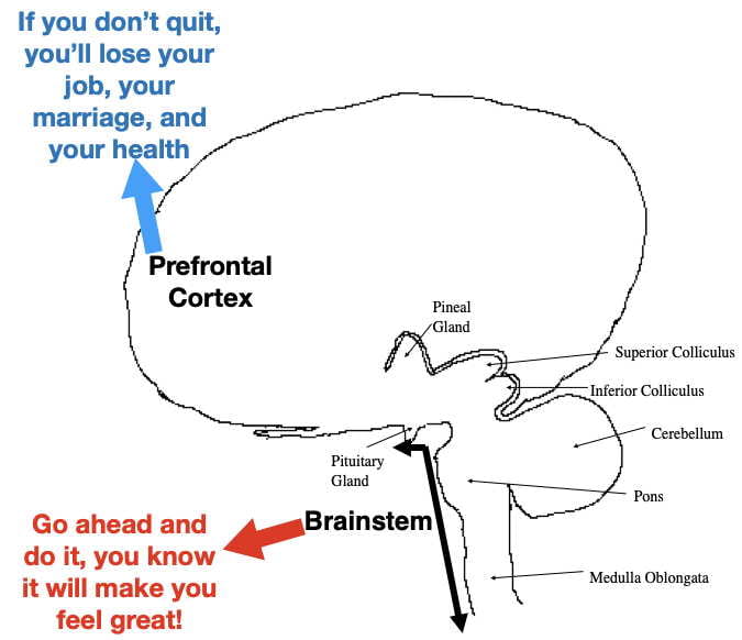
While the highly modified mammalian forebrain (i.e. cortex, limbic system, basal ganglia, and the fiber tracts that interconnect them) expanded behavioral flexibility and promoted broader adaptation, the more primitive brainstem remained tightly linked to unconscious homeostatic processes such as breathing, blood flow, body temperature regulation, etc.  In the unusual situation where higher and lower brain areas would be in conflict regarding these critical processes, the lower brain areas should normally win out.  For example, it’s virtually impossible to commit suicide by consciously holding your breath.  No matter how hard you try, the unconscious breathing centers in the medulla (lowest part of the brain) will make you start breathing again.  (Hopefully nobody reading this proves me wrong!)  It is also instructive to look at the effects of brain damage.  Because the medulla is in charge of homeostatic processes critical to life, damage to the primitive medulla is much more likely to be lethal than damage to the much more complicated cortex.

Conflicts arising from independent processing in different parts of the brain.

While independent processing in different parts of the brain usually works together, it is possible to have conflicts.  Some of the best examples are found in split-brain preparations where the corpus callosum (the fiber tract that connects the two cortical hemispheres) has been cut (typically to treat epilepsy), causing the 2 hemispheres to be disconnected in their functioning.  When this happens, the two hemispheres are less able to coordinate their processing and occasionally bizarre outcomes can occur where one hemisphere will try to produce one set of motor responses while the other hemisphere will try to produce another.  More important to the addiction argument would be conflicts between higher and lower brain areas.  While higher and lower brain areas also usually work together, conflicting processing should be possible here as well.

A good example of independent processing by higher and lower brain areas is the brain’s processing of visual information.  As seen in the figure below, our conscious visual experience is begun by processing occurring in the primary visual cortex of the occipital lobe.  However, a lower part of the human brain (the superior colliculus in the midbrain) is also independently processing visual input, but below the level of conscious awareness.  In fact, for vertebrates that lack a neocortex (such as reptiles and birds), the optic tectum (equivalent to the human superior colliculus) is the primary visual processing center.

Figure 2: A cross section schematic of the human brain showing the visual pathways and 2 visual processing areas in red. The lateral geniculate nucleus (LGN) is a relay station where optic nerve fibers must first synapse before sending the information up to the primary visual cortex in the occipital lobe via a fiber tract called the optic radiation.  In contrast, optic nerve fibers go directly to the superior colliculus

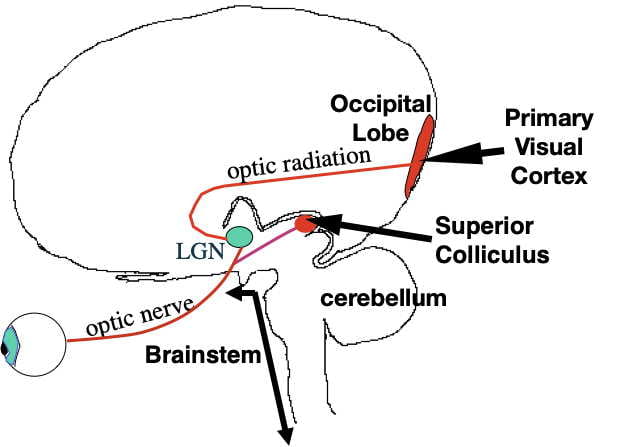
Visual processing by the superior colliculus is thought by some to to explain another bizarre phenomenon in humans known as “blindsight”.  Blindsight occurs following brain damage to the primary visual cortex in the occipital lobe of the cortex (necessary for conscious visual processing).  Such a person would be diagnosed as blind by an ophthalmologist. However, if the eyes, the optic nerve, and visual pathways into the lower parts of the brain including the superior colliculus are undamaged,  such a “blind” person can still behaviorally respond to certain types of visual input.  However, when asked what caused the response and why they responded, the person cannot give a good explanation.  A commonly accepted explanation is that the visual input is being processed by the superior colliculus operating below the level of conscious awareness.

While both the human visual cortex and superior colliculus normally work together, their independent visual processing  is thought to serve different purposes.  While the much more sophisticated cortical processing influences conscious thought and behavior, the simpler superior colliculus processing may be important for quick reflexive responses to visual input.   We do know that the human superior colliculus does process visual input to influence several different reflexive eye and head movements known to facilitate vision.  However, whether the superior colliculus underlies blindsight is based more upon indirect evidence.  Like the primary visual cortex, a retinotopic map exists in the superior colliculus where specific locations in the superior colliculus correspond to particular locations in the retina.   The relationship suggests that the human superior colliculus might have the neural machinery to reconstruct some representation of what the person is seeing. The superior colliculus also is connected to the spinal cord by a fiber tract (the tectospinal pathway) that could mediate quick output to human body muscles as it does in birds and reptiles.  This role also makes anatomical sense because the superior colliculus both receives visual information sooner and, can reflect it back out to muscles faster than the cortex, allowing for quick (but unconscious) visual reflexes.

The point of this digression is that while different parts of the brain normally work together, independent processing of similar information does occur at different brain levels.  And should conflicts arise concerning issues important to survival and reproductive fitness, the lower brain areas should sometimes have priority.  And few things are more important to survival than quickly identifying and responding to rewarding (and aversive) stimuli.

So where is the addiction circuitry? 

I offer the hypothesis (not original with me) that addiction may be influenced by memory circuitry somewhere in the unconscious parts of the brain relatively immune to the influence of conscious learning.  Exactly where is a question I won’t try to answer.   However, I would suggest that the lower this circuitry is in the brain, the more resistant it would be.  I will now weasel out of the situation by hoping that future research will be able to address this very important question.  I will also leave it to persons smarter than me to figure out what, if any, implications this could have for addiction treatment.

As I pointed out in the first post on addiction, the prevailing school of thought is that addiction can be accounted for by dysfunctions in the reward circuitry as well as in the prefrontal cortex and amygdala.  I am not discounting the important role these forebrain structures play, particularly in encoding the initial conscious reward expectancies.  However, I am suggesting as addiction proceeds, the forebrain dysfunctions (particularly in the prefrontal cortex) may contribute even more by impairing the organized conscious thought processes necessary to override powerful primeval reward expectancies unconsciously encoded the brainstem.

Figure 3. A cartoon showing the proposed conflict between conscious and unconscious parts of the brain in a drug addict

The reality is that we don’t really completely understand what encodes and maintains the need to engage in this highly compulsive, long-term, out-of-control behavior and all explanations (including the one presented here) should be taken with a grain of salt.

Sorry to take so long to reach such an unsatisfying conclusion.  🤷‍♂️But…….that’s the way science sometimes goes!  Still a lot to learn!

To learn more.

Advocat, Comaty & Julien (2019).  Chapter 4: Epidemiology and neurobiology of addiction.  In Julien’s Primer of Drug Action. Thirteenth Edition. Worth Publishers. 267-295.  (A good textbook that I last used in my teaching.  many references to primary literature at the end of this chapter)

Charles P. O’Brian.  Drug use disorders and addiction.  Chapter 24 in Goodman and Gilman’s The pharmacological basis of therapeutics.  McGraw-Hill Publishers.  (written for medical professionals so fairly technical)

I also refer you to Wikipedia for information on addiction, split-brain preparations, blindsight, and consciousness.  The Wikipedia entries also provide citations to the primary literature.

John Nyby:wordpress.lehigh.edu/jgn2


Addiction I: The Role of Reward in Addiction

Introduction

The U.S. is clearly a nation of drug users.   On a per capita basis, our combined legal and illicit drug use exceeds that of any other country.  And for those drugs that are abused (either illegal or prescription), a certain percentage of users become addicted.  In 2015, drug overdose deaths finally managed to surpass automobile accidents as the leading cause of preventable deaths (with opioids being the main culprit).   Over ½ trillion dollars are spent each year combating U.S. drug abuse, with harder-to-measure social and interpersonal costs being equally serious.  Despite governmental efforts, drug abuse and addiction are  societal problems we have yet to get a handle on.

A major problem in dealing with drug addiction is how little we are really sure about.  Like many complex biological and psychological phenomena, it is difficult to come up with a precise definition of addiction that everyone accepts.  Although the brain is definitely involved, there remain mysteries concerning exactly how it is involved.  While addiction clearly has a genetic component, it’s not always apparent how genes provide this susceptibility.  And while many treatments can provide some relief, they often function more as temporary “band-aids” than as long-term “cures”.

This blog posting is the first in a series of four. In this posting, I begin by addressing definitional issues and how scientific thinking about the causes of addiction have changed over the years.  In the second, I get nerdy  and speculate about the critical brain circuitry and where this circuitry might be located (hopefully in an understandable way).  In the third posting, I briefly explore genetic and environmental factors related to addiction.  And in the final posting, I present an overview of some different pharmacological treatments to help addicts cope.

Definition of Addiction.

There are probably as many definitions of addiction as there are experts.  At the same time, there are three addiction characteristics upon which most would agree.  1. An addict has uncontrollable cravings to take the drug.  2.  An addict will continue to take the drug despite experiencing negative consequences.  3.  Once addicted, an addict has great difficulty stopping.

A major problem in defining addiction is that it exists on a continuum. While some drug users are easily identified as addicts and others not, for individuals somewhere in the middle, classification is not always clear.  The question is, where do you draw the line?  I find the definition of Burgess and Shaffer in 1984 both instructive and amusing.  They define addicts as “certain individuals who use certain substances in certain ways thought at certain times to be unacceptable by certain other individuals for reasons both certain and uncertain.”

The Diagnostic and Statistical Manual of the American Psychiatric Association (DSM V) has attempted to resolve the issue, not by defining addiction per se, but rather by operationally defining a disease called substance abuse disorder (roughly comparable to addiction).   To do so, DSM V identifies 11 possible problems that a regular drug user might have.   Different degrees of this disease (mild, moderate, and severe) are then defined by how many of these 11 problems occur.

However, according to DSM V, regular drug use, by itself, does not qualify a person as having substance abuse disorder.  The person must have at least 2 of the cited problems for the mild form of the disorder.  Although unlikely, it is theoretically possible for a chronic drug user to not qualify, an outcome some drug experts find problematic.

Despite definitional problems, the term “addiction” continues to be heavily used by the public as well as by professionals.  While addiction is difficult to define precisely, most of us know it when we see it.

Tolerance and withdrawal

The original idea of how people become addicted was explained by the well-established pharmacological principles of tolerance and withdrawal.  When you first take an addictive drug, body and mental functioning are often disturbed.  However, with continued use, the body makes homeostatic compensations to reduce the disturbance and allow the user to function more normally in the presence of the drug.   As this tolerance to the drug become established, it also causes a given dosage of drug to become less rewarding.   To continue to obtain the desired reward, larger and larger drug dosages must be taken.

Two major types of compensation underly tolerance.  For example, many addictive drugs exert their effects by binding specific neurotransmitter or neurohormone receptors embedded in neuron membranes in the brain.  Under conditions of continued binding, the brain compensates by downregulating (decreasing the number of) these receptors, which makes the drug less effective.  Another type of compensation occurs mainly in the liver where most drugs are enzymatically deactivated and prepared for excretion.  The liver compensates by upregulating the appropriate enzyme(s), further contributing to the drug’s loss of effectiveness.  This “double whammy” causes tolerance, resulting in more drug being required to get the desired effect.

In addition, once the body has established a new homeostatic set-point to allow for functioning in the presence of the drug, the drug must then be present in the body allow for proper body functioning.  If the drug is discontinued, body homeostasis is again thrown out of whack and the person experiences withdrawal.

Withdrawal symptoms are, in many ways, the opposite of the drug effects.  Since addictive drugs make you feel good, withdrawal makes you feel bad.  During withdrawal, the brain’s autonomic regulation of body physiology begins to malfunction.    So, until your body can re-establish its pre-drug homeostasis, you experience symptoms of withdrawal.  The intensity of withdrawal and how long it takes, depends on the drug and the degree of tolerance achieved.   Sometimes it can take a month or more of drug abstinence for withdrawal symptoms to subside.

Although withdrawal symptoms vary somewhat from drug to drug, in all cases withdrawal results in a dysphoric, anxious, hyperactive, and unhealthy state.  And in all cases, a very easy way to make the unpleasant withdrawal symptoms go away is to take the drug.  According to this idea, once you’ve reached the stage where significant withdrawal can occur, you are now addicted.

This perspective of addiction was likely developed from studying alcoholics, and perhaps heroin addicts, where it seemed to have some utility.  While withdrawal occurs to all drugs of abuse, alcohol withdrawal generally has the most serious consequences.  For example, the withdrawal from alcohol by a chronic alcoholic is called delirium tremens (DT’s).  It is not only very unpleasant, in extreme cases, particularly in older, long-term alcoholics, it can be fatal (usually through heart attack or stroke).  However, the DT’s are quickly alleviated by taking a drink of alcohol.  Thus, an alcoholic was viewed as being addicted in order to prevent the DT’s.

However, the tolerance/withdrawal explanation has some problems.  One problem is that after withdrawal is complete and brain receptors and body physiology have pretty much returned to normal, all withdrawal symptoms subside except for one:  the compulsive desire to take the drug.  Some experts argue that once addicted, you are addicted for life.

Another problem is that some non-addictive drugs  (such as antidepressants) can cause marked tolerance and decidedly unpleasant withdrawal.  But after withdrawal is complete, they do not result in an addictive desire to take the drug.  It’s also problematic that marijuana, a drug with addiction potential, does not have withdrawal symptoms for many users.  (Marijuana users do develop tolerance. However, during abstinence, the very gradual release of THC into the blood from its storage in body fat produces a unique “built-in” tapering effect that minimizes withdrawal symptoms.  In fact, when addicts are trying to quit other addictive drugs, gradually reducing the dosage is sometimes used to help them cope with withdrawal.)

While the desire to avoid withdrawal can certainly influence an addict’s day-to-day drug use, the hard-to-control desires that remain after withdrawal is complete must be due to something else.

Brain Reward Circuitry

One of the great discoveries of modern neuroscience is that rewarding stimuli (e.g. food, drink, warmth, sex, social interactions, and drugs) are thought by many neuroscientists to be experienced as pleasurable and rewarding through activation of a common reward circuitry in the brain.  Critically important to this circuitry are ventral tegmental neurons in the midbrain that release dopamine into the nucleus accumbens in the forebrain via part of an ascending fiber tract called the mesolimbic/mesocortical pathway (See Figure 1 below).  Other parts of the mesolimbic/mesocortical pathway (not shown below) deliver dopamine from ventral tegmental neurons to parts of the amygdala (important in emotional responses), prefrontal cortex (the executive part of the cortex that makes conscious decisions about what you should be doing and when you should be doing it) and, to some degree, the hippocampus (important in memory formation).  All addictive drugs , either directly, or indirectly, are now thought to promote this dopamine release in varying degrees.  Moreover, non-addictive neuroactive drugs  (like most antidepressants and cannibidiol (CBD) ) do not.

Figure 1: A schematic cross section of the human brain showing the reward circuitry in red.  The neurons of this circuitry have their cell bodies in the ventral tegmental area and their axons project up to the nucleus accumbens where they secrete dopamine under conditions of reward.  These axons are part of a larger ascending fiber tract called the mesolimbic/mesocortical pathway.

There are changes in the areas that receive dopamine input as a result of chronic drug use that last for some time.  For example, in regular cocaine users dopaminergic synapses in the nucleus accumbens are strengthened (making the nucleus accumbens more sensitive to dopamine).  There is also a strengthening of glutaminergic input into the ventral tegmental area, altering the ability of other brain areas (such as the prefrontal cortex and amygdala) to activate the reward circuitry.  Accompanying changes in the reward circuitry are also changes in the functioning of the prefrontal cortex and amygdala.

These dopamine-related changes have been proposed to underly the neurological basis of addiction.  While they are almost certainly involved in the development of an addiction, they may not be sufficient, by themselves, to explain long-term addiction.  The problem is that the reward circuitry, and associated areas, also influence many other areas of the brain.  Consequently, important changes underlying long-term compulsive drug use may actually occur elsewhere as well.

In the next post, I present an idea about what that might entail.

John Nyby:wordpress.lehigh.edu/jgn2


Your Brain On THC, The Psychoactive Ingredient In Marijuana

Introduction.

There is still a lot that we don’t know about THC, the psychoactive phytocannabinoid in marijuana.  A major reason is that marijuana is illegal in many countries.  Marijuana’s federal illegality in the United States (although many states beg to differ) and a corresponding lack of government-sponsored funding have certainly discouraged research here in the United States.

But with the recent complete legalization in Canada and South Africa, many more laboratories around the world have begun to engage.  I anticipate changes in US laws in the near future further contributing to significant discoveries over the next several decades.

After some background on cannabis plants, I try to give you an understanding of how THC interacts with neurons in your brain to ultimately produce its many effects upon physiology and behavior.   I will not be describing those many effects as they have been covered extensively elsewhere.

A disclaimer before starting.  My explanations and speculations represent my current understanding.  However, cannabis research is advancing quickly, and it is possible that some of my explanations will need to be altered or modified in the future in terms of new research.

Three different cannabis plants.

THC is a phytocannabinoid unique to the 3 different varieties of cannabis plants (Cannabis sativa, Cannibis indica, and Cannabis ruderalis).  Whether taxonomists classify them as separate species or subspecies depends upon which taxonomist you talk to.  However, they can all interbreed.   All three had their origins as wild plants indigenous to different parts of Asia, although only sativa and indica have been domesticated.  In fact, many, if not most, domesticated marijuana plants are now hybrids of sativa and indica.  The current world-wide use of domesticated marijuana was initiated no doubt via trade routes in Asia, Europe, Africa, and eventually the remainder of the world.  One consequence, is that cannabis  is now growing wild around the world, including the United States.

Number of phytocannabinoids.

THC is only one of many phytocannabinoids found in cannabis plants.  When I began teaching a neuropharmacology course about 15 years ago there were 60 known phytocannabinoids.  However, I recently read the known number has grown to 150.  I suspect the final tally has yet to be determined.   At the same time, many phytocannabinoids occur in negligible amounts and may be either precursors or metabolites of the more prevalent ones and probably make a negligible contribution to marijuana’s effects.  Most of the attention so far has been on tetrahydrocannabinol (THC) for its psychoactive effects and Cannabidiol (CBD) for its medical benefits.  In this blog, I will focus on THC.

Before getting into the details of THC action in the brain, I present some additional background.

Why do cannabis plants make THC and other cannabinoids?

Phytocannabinoids did not evolve to serve the recreational, medicinal or religious needs of humans.  That is an unintended consequence.  Rather, phytocannabinoids exist to provide benefits to the marijuana plant itself.   So….. what are the benefits?   Given the large number of phytocannabinoids, a variety of purposes are likely served.

To begin to understand function, it is instructive to look at the how similar molecules benefit other source plants.  For example, the alkaloids made by many plants (e.g. cocaine, nicotine, strychnine, caffeine, morphine, pilocarpine, atropine, methamphetamine, mescaline, ephedrine, and tryptamine) are also psychoactive drugs that alter neurotransmission and are also used by humans for recreational and medical use.  In high dosages, some are even lethal.  Although vertebrates, including humans, are clearly affected, insects are even more susceptible.  Although THC is not classified as an alkaloid (because it lacks nitrogen atoms), I would suggest that THC may have analogous functions.

For example, many of the alkaloids mentioned above serve as natural “insecticides” to minimize the likelihood of the host plant being eaten.  In fact, many years ago, tobacco extracts were employed as an insecticide.  Unfortunately, concentrated nicotine can also be highly poisonous to humans.   However, chemists were able to alter nicotine’s chemical structure to make it less poisonous to humans while, at the same time, retaining potent insecticide properties.  The resulting “neonicotinoid” insecticides subsequently replaced nicotine and became very popular around the world.  While still in use by farmers, several of these insecticides are currently banned by the European Union and restricted by several American states because they are considered a contributing factor in the world-wide decline in honeybee populations.  It seems reasonable that THC similarly protects the cannabis plant from being eaten by causing nervous system dysfunction of the animal that eats it.

Yet another way that alkaloids protect their host plant is by tasting bitter.  In fact, the benefit of being able to reject alkaloid-containing plant food was instrumental in the evolution of animal bitter taste receptors.  While we may think of bitterness as a unitary taste quality, many different “bitter” receptors exist to detect the wide range of alkaloid molecules that taste bitter.  In fact, animals have evolved many more genes to encode bitter receptors than for sweet, sour, or salty tastes.  The large number of genes provides indirect evidence for the importance of this taste quality. Although cannnabinoids themselves are not as bitter tasting, the plant does have distinctive taste and odor qualities from its terpenes as well as its cannabinoids that could serve similar deterrent functions, particularly to indigenous animals that provided the greatest threat over evolutionary time.

Alkaloids also serve other functions as well.  For example, caffeine released into the soil by coffee seedlings inhibits the germination of nearby seedlings, thereby reducing competition for resources.  This allows the seedlings to better spread themselves out to optimize growth and development.  As the field of cannabis research becomes more mature, I anticipate that additional phytocannabinoid functions will become apparent as well.

However, once domesticated, a new set of evolutionary selection pressures arose.  Human growers began subjecting domesticated cannabis to intense artificial selection to emphasize the phytocannabinoid profiles that contribute to human recreational and medical use.  This selection pressure has clearly impacted the relative concentrations.

For example (as seen below), the US Drug Enforcement Administration, in conjunction with the University of Mississippi‘s Potency Measuring Project, found that THC concentration in confiscated marijuana cigarettes increased dramatically after 1978.   Today’s marijuana is definitely not your parents and grandparents’ marijuana!

Table 1. The average concentration of THC in marijuana cigarettes over a period of 30 years.

Pretty interesting that plants figured out how to protect themselves from being eaten by evolving chemicals that mess around with the brains of animals!  Just as interesting is that humans figured out how to use these “poisons” for recreational, medical and religious purposes.

Endocannabinoids.

Just as marijuana plants make multiple phytocannabinoids, vertebrates, including humans, produce multiple endocannabinoids.  Endocannabinoids are the naturally occurring chemical messengers in our bodies that interact with the 2 classes of cannabinoid receptors (CB-1 and CB-2).  Although both receptors exist inside and outside the brain, CB-1 is important inside the brain while CB-2 is more important outside the brain.  The phytocannabinoid of interest, THC, exerts its psychoactive effects primarily by interacting with CB-1 receptors in the brain.

While as many as 7 different endocannabinoids have been identified, the 2 most studied are anandamide (which is a partial CB-1 agonist like THC) and 2-AG (a full CB-1 agonist).  Like neurotransmitters, endocannabinoids can be synthesized by neurons, travel across synapses, and bind to receptors on the other side of the synapse.  However, endocannabinoids differ from conventional neurotransmitters in that they move in the opposite direction, from the postsynaptic membrane back to receptors embedded in the presynaptic membrane.  Consequently, endocannabinoids are sometimes called retrograde neurotransmitters or retrograde messengers.  Below you can see a schematic that will hopefully allow you to understand the microanatomy involved.

Figure 1. A synapse showing the sequence of events from endocannabinoid synthesis, release from the postsynaptic membrane, traveling backwards across the synapse, CB-1 receptor binding, and ultimately inhibition of anterograde neurotransmitter release. After completing their job, endocannabinoids are taken inside neurons and destroyed.

The molecular structures of the different endocannabinoids, while molecularly similar to each other  (all are derived from  arachidonic acid), bear little resemblance to THC.   However, some part of THC and endocannabinoid structures must be shaped similarly in order that they all fit in a “lock and key” fashion into the same receptor binding site.  The very different molecular structures of THC, Anandamide and 2-AG are seen below.

Figure 2. The molecular structure of THC versus the 2 most studied endocannabinoids, anandamide and 2-AG
Figure 2.  The molecular structures of THC, Anandamide, and 2-AG.  Despite the endocannabinoids being very different from THC, all three molecules bind and activate the CB-1 receptor.

Function of the endocannabinoids inside the brain.

In the human brain the endocannabinoids are thought to be part of a protective system that prolongs the functioning of “over-worked” neurons.  It turns out that each neuron has a finite amount of neurotransmitter storage.  So, when a neuron becomes hyperactive for an extended period of time, it can potentially deplete its neurotransmitter storage and cease to function properly.  However, endocannabinoid secretion serves to minimize this outcome.

The way this works is the postsynaptic neuron can sense abnormally high levels of  neurotransmitter release which causes endocannabinoid release backwards across the synapse.  The endocannabinoids then bind to CB-1 receptors in the presynaptic membrane which in turn slows the release of the anterograde neurotransmitter.

This negative feedback prolongs the ability of the presynaptic neuron to function (albeit at a lower level) reducing the likelihood that the neuron will drive its neurotransmitter storage to exhaustion.  Virtually all brain synapses outside the brain stem (regardless of neurotransmitter type) are thought to have this protective mechanism.  In fact, some scientists think the CB-1 receptor may be the most common receptor in the brain.  This protection is analogous to a governor on a school bus that shuts off gasoline to the engine at a predetermined speed to prevent the bus driver from going too fast.

The endocannabinoid system normally just sits there quiescent until a particular part of the brain becomes hyperactive and is activated on an as-needed basis.  When activated, neurotransmission is slowed ONLY in the hyperactive part of the brain.  So, the effects are normally localized to the hyperactive brain area, with most of the brain being unaffected.

So how does THC work in the brain?

In contrast, THC administered by smoking or eating marijuana is not localized, but rather floods the whole brain. This pattern of CB-1 receptor activation is obviously very different from that of the endocannabinoids.  The resulting interactions throughout the brain areas affected give rise to the many effects of THC on sensory, motor, emotional, and cognitive functioning.

As I mentioned earlier, THC is only a partial agonist for the CB-1 receptor.  When you flood the brain with THC, only around 20% of the CB-1 receptors are bound at any given instant of time.  This is in contrast to anandamide, a more potent partial agonist that binds around 50%, and 2-AG which is a full agonist binding virtually all the receptors.  Thus, the profound psychoactive effects of THC are not because it is a powerful activator of CB-1 receptors but rather because there are so many CB-1 receptors in so many different parts of the brain that binding only a small fraction can have profound effects.

However, unlike some alkaloids that are lethal in high dosages, no one to my knowledge has ever died from a marijuana overdose.  The reason is that, unlike the receptors affected by alkaloids, there are few or no CB-1 receptors in the brain stem.  Thus, the areas of the brain that control processes essential to life such as breathing and heart functioning are only minimally affected by THC.

Concluding remarks.

I’ve obviously left out a lot of information, so I encourage you to pursue this and related topics on your own.  As mentioned earlier, there is also great interest in cannabidiol (CBD) which accounts for some of the more profound medical benefits of cannabis.  CBD is not psychoactive and does not seem to be an agonist for either the CB-1 or CB-2 receptor, although it may partially block the CB-1 receptor.  In fact, relatively little is known about how CBD produces its effects.  As more becomes known about its mechanism of action, perhaps I can write a blog on this important phytocannabinoid.

Some other phytocannabinoids that are beginning to receive attention for possible medicinal properties are cannabigerol (CBG), tetrahydrocannabinolic acid (THCA) and tetra hydrocannabivarin (THCV).  However, little scientific attention has been paid to most of the phytocannabinoids.   While the rarer phytocannabinoids may make only a negligible contribution to marijuana’s effects under normal circumstances, when isolated and concentrated (or synthesized) they may turn out to have profound effects as well.  It also seems likely that some marijuana effects are influenced by interactions among phytocannabinoids.  For example, we know that CBD both mutes and prolongs the psychoactive effects of THC.

And finally, phytocannabinoids likely have significant hormone-like effects outside the brain. The tissues of the digestive system, liver, kidney, and heart, as well as immune cells all have cannabinoid receptors.   In fact, the cannabinoid hyperemesis syndrome sometimes seen in long-term marijuana users is thought to be caused by phytocannabinoid binding outside the brain.   Hormone-like effects outside the brain definitely merit serious study.

To learn more.

Advocat, Comaty & Julien (2019).  Chapter 9: Cannabis.  In Julien’s Primer of Drug Action. Thirteenth Edition. Worth Publishers. 267-295.  (many references to primary literature at the end of this chapter)

The Health Effects of Cannabis and Cannabinoids: The Current State of Evidence and Recommendations for Research (2017)By National Academies of Sciences, Engineering, and Medicine, Health and Medicine Division, Board on Population Health and Public Health Practice, Committee on the Health Effects of Marijuana: An Evidence Review and Research Agenda.  The National Academies Press. (a very comprehensive analysis of cannabis research with an extensive bibliography)

John Nyby:wordpress.lehigh.edu/jgn2